For the safety of patients and staff, please wear a mask if you have cold or flu-like symptoms.
Request an Appointment
Understanding X-rays: how they work and what to expect

If you have ever been handed a requisition for an X-ray and felt a flash of worry about radiation or what the procedure actually involves, you are not alone. Many Canadians feel uncertain about imaging exams, even routine ones. The good news is that X-rays are one of the safest and most well-understood diagnostic tools available, and knowing what to expect makes the entire experience far less stressful. This guide walks you through how X-rays work, what types exist, how safety is managed, and what your visit to an Ottawa imaging clinic will actually look like.
Key Takeaways
| Point | Details |
|---|---|
| X-rays are safe and essential | Modern X-rays use low doses of radiation and are invaluable for diagnosing many health problems. |
| Different types for different needs | From plain film to fluoroscopy and CT, each X-ray technique serves unique diagnostic purposes. |
| Balance benefits and risks | The health benefits of accurate diagnosis far outweigh the small risks—safety protocols are built into every exam. |
| Ottawa uses expert criteria | Doctors in Ottawa follow evidence-based criteria to ensure X-rays are only used when necessary. |
How do X-rays work?
X-rays are a form of invisible, high-energy electromagnetic radiation. When a beam of X-rays passes through your body, different tissues absorb different amounts of that energy. Dense structures like bone absorb a great deal, while softer tissues like muscle and fat let more radiation pass through. This difference in absorption is what creates the contrast you see on an X-ray image.

According to MedlinePlus, X-rays direct radiation through the body onto a detector or film, where denser tissues absorb more radiation and create a contrasting shadow image. Bone appears white or light grey, while air-filled spaces like the lungs appear dark. Soft tissues fall somewhere in between, showing up as various shades of grey.
Here is a simple breakdown of what happens during a standard X-ray:
- You are positioned by a trained technologist, either standing, sitting, or lying down, depending on the area being examined.
- A small amount of radiation is directed through the targeted body part for a fraction of a second.
- The detector or digital sensor captures the image almost instantly.
- The image is sent electronically to a radiologist (a physician who specialises in reading diagnostic images) for interpretation.
- Results are typically available within 24 to 48 hours at most Ottawa clinics.
You can learn more about X-ray technology explained and what to expect during your appointment. The entire process is painless and, for most standard exams, takes only a few minutes from start to finish.
With the basics established, let’s look at the different types of X-ray procedures and what sets them apart.
Types of X-ray procedures and their uses
Not all X-rays are the same. The type your doctor orders depends on what they are trying to see and why. As outlined by the Merck Manuals, imaging variations include plain radiography for bones and lungs, fluoroscopy for real-time motion imaging, angiography for blood vessels using contrast dye, barium studies for the gastrointestinal tract, and CT scans for detailed cross-sectional images.
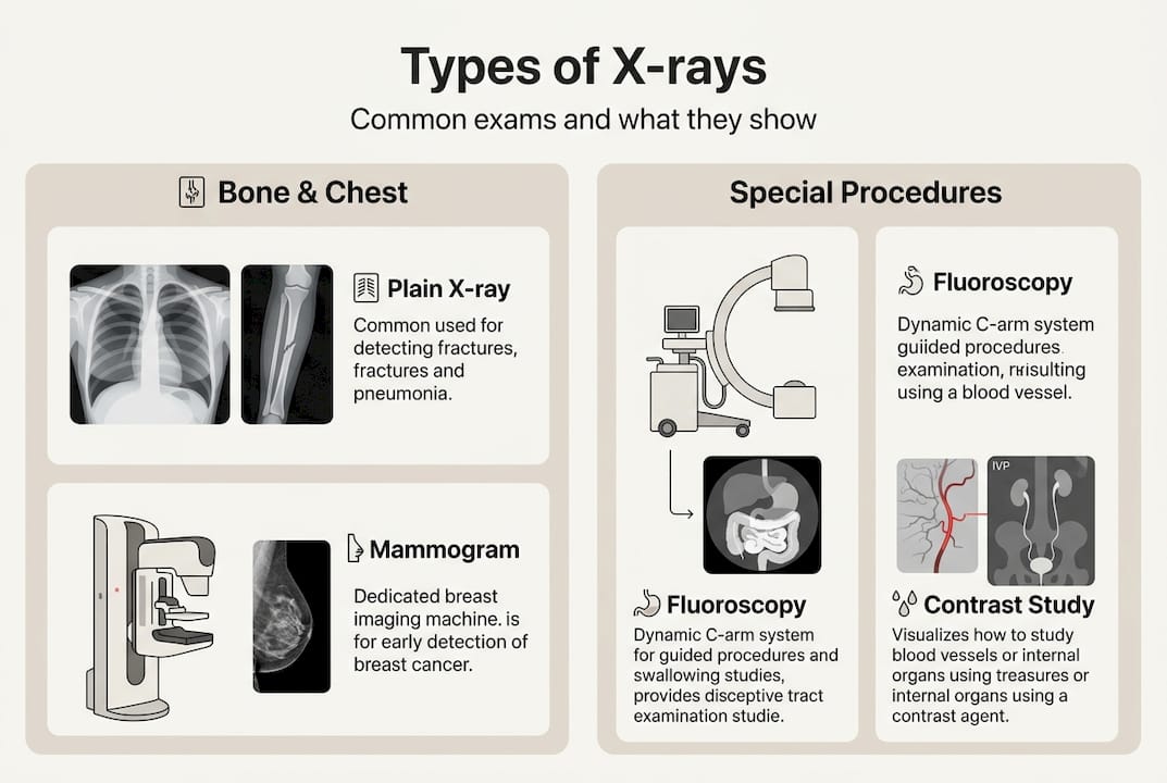
| Type | What it shows | Common uses |
|---|---|---|
| Plain radiography | Bones, lungs, teeth | Fractures, pneumonia, dental issues |
| Fluoroscopy | Moving structures in real time | Swallowing studies, joint injections |
| Angiography | Blood vessels with contrast | Blocked arteries, vascular conditions |
| Barium studies | Digestive tract | Ulcers, bowel conditions |
| CT scan | Detailed cross-sections | Cancer staging, trauma assessment |
Your doctor chooses the right type based on your symptoms, medical history, and what information they need to make a diagnosis. For example, a simple chest X-ray is ideal for checking lung health, while a CT scan provides far more detail for complex conditions.
Understanding the difference between comparing X-ray and fluoroscopy can also help you feel more prepared when your doctor explains their recommendation.
Pro Tip: If your doctor orders a specific type of X-ray and you are unsure why, ask them to explain which type it is and what they are looking for. A clear understanding of the purpose helps reduce anxiety before your appointment.
Now that you know the various types of X-ray exams, it’s important to understand the safety considerations involved.
Understanding X-ray safety: risks and dosages
Radiation exposure is the concern most patients raise first. It helps to put the numbers in perspective. Standard X-ray doses are low, with a chest X-ray delivering between 0.02 and 0.1 millisieverts (mSv), which is roughly equivalent to 2 to 10 days of natural background radiation from the environment. More advanced procedures like CT scans involve higher doses, sometimes several mSv, which is why they are used only when clinically necessary.
Here is a comparison of common X-ray doses:
| Exam type | Approximate dose (mSv) | Equivalent background radiation |
|---|---|---|
| Chest X-ray (PA view) | 0.02 to 0.1 | 2 to 10 days |
| Lumbar spine X-ray | 1.5 | 6 months |
| CT abdomen/pelvis | 8 to 15 | 3 to 5 years |
| Dental X-ray (bitewing) | 0.005 | Less than 1 day |
Canadian imaging clinics follow the ALARA principle, which stands for “As Low As Reasonably Achievable.” This means technologists use the minimum radiation dose needed to produce a clear, diagnostic-quality image. You can read more about the number of safe X-rays over a lifetime and how cumulative exposure is tracked.
The MSD Manuals note that risks for single exams are minimal, but the ALARA principle applies, particularly for pregnant patients and children, where shielding and protocol adjustments are standard practice. The benefits of accurate diagnosis almost always outweigh the small risk from a single exam.
One important advancement worth knowing about is digital versus traditional X-rays. Digital systems produce high-quality images at lower radiation doses compared to older film-based technology, and they allow images to be shared instantly with your care team.
Pro Tip: If you are pregnant or think you might be, always tell the technologist before your exam. They will take appropriate precautions, and in some cases, may suggest an alternative imaging method. Strategies for minimising radiation exposure are always a priority at accredited clinics.
Safety is a top concern, but what if special circumstances or rare reactions arise? Let’s look at some edge cases and real-world considerations.
Special considerations: contrast agents, edge cases, and new technology
Most X-rays are straightforward, but some situations require extra attention. The NIBIB outlines that edge cases include rare allergic reactions to contrast agents like barium or iodine, higher doses associated with interventional fluoroscopy, and the need for optimised protocols in paediatric patients.
Here is what you should know about the most common edge cases:
- Contrast agent reactions: Barium and iodine-based contrast agents are used to highlight specific structures. Allergic reactions are rare but possible. Clinics monitor patients closely and have protocols in place to respond quickly.
- Paediatric adjustments: Children are more sensitive to radiation, so technologists use lower doses and more targeted shielding. Protocols are specifically designed for younger patients.
- Interventional fluoroscopy: Procedures like guided injections or catheter placements involve longer exposure times. Radiologists carefully weigh the risks and benefits beforehand.
- Digital advancements: According to UK national dose surveys, digital detectors reduce dose compared to traditional film, improving both safety and image quality.
Important: Always inform your care team about any known allergies, previous reactions to contrast agents, or existing health conditions before your exam. This information helps them prepare the safest possible experience for you.
The advantages of digital imaging are significant, including faster processing, better image clarity, and reduced radiation exposure. If you have concerns about contrast agents, you can also learn more about contrast allergies explained and how they are managed.
All these factors show that X-ray diagnostics are more nuanced than most people assume. But how do you know when to get an X-ray and what happens day-to-day in Ottawa clinics?
When X-rays are recommended: Ottawa rules and practical application
One of the most useful things to know is that doctors in Ottawa and across Canada use established clinical guidelines to decide whether an X-ray is truly necessary. For example, the Ottawa Ankle Rules and Ottawa Knee Rules are evidence-based criteria that help emergency physicians determine whether imaging is needed after an injury, reducing unnecessary exams.
Understanding when to get an X-ray can save you time and reduce unnecessary exposure. The RSNA’s appropriate utilisation guidelines outline when imaging is genuinely warranted versus when watchful waiting or alternative tests are more appropriate.
Here are some practical tips for your first visit to an Ottawa imaging centre:
- Bring your requisition form from your doctor. Most clinics require it before the exam.
- Wear comfortable clothing without metal fasteners, as you may need to remove items near the area being imaged.
- Arrive a few minutes early to complete any necessary paperwork.
- Ask questions freely. The technologist is there to help you feel comfortable and informed.
- Discuss alternatives with your doctor if you have concerns. Ultrasound and MRI do not use ionising radiation and are sometimes equally effective for certain conditions.
Pro Tip: Before your appointment, write down any questions you have for the technologist or radiologist. Asking about the purpose of the exam, the estimated wait time for results, and any follow-up steps puts you in control of your care.
With practical advice in hand, let’s turn to what most guides don’t tell you about getting the most value from your diagnostic experience.
What most guides miss about X-ray diagnostics
Most articles focus entirely on how X-rays work and stop there. What they rarely address is the importance of questioning whether an X-ray is the right tool for your situation in the first place. More imaging is not always better. Appropriateness criteria exist precisely because unnecessary exams add cumulative dose, cost, and sometimes anxiety without improving outcomes.
We believe the most empowered patients are those who understand knowing when X-rays are truly necessary and feel comfortable asking their care team about alternatives. Technology improvements like digital detectors and AI-assisted image analysis are making X-rays safer and more accurate, but the most important factor remains a collaborative relationship between you and your radiology team. Ask questions. Share your history. Advocate for the exam that genuinely serves your health.
Where to get X-rays and advanced imaging in Ottawa
If you or someone you care for needs an X-ray in Ottawa, Canadian Diagnostic Network (CDN) offers convenient access across more than 20 clinic locations. Our experienced team of radiologists and technologists delivers accurate results, typically within 24 to 48 hours. For urgent needs, our walk-in urgent care imaging service means you do not always need to wait for a scheduled appointment. You can also visit Broadview Imaging Ottawa for full-service diagnostic imaging close to home. We are here to make your experience as smooth and reassuring as possible.
Frequently asked questions
Are X-rays safe for children and pregnant women?
Standard X-ray exams carry low risk, but paediatric protocols are optimised for lower doses and pregnant women should only have X-rays when clinically necessary, with appropriate shielding used in all cases.
How long does an X-ray take and does it hurt?
Most X-rays take just a few minutes from start to finish. As MedlinePlus confirms, X-ray beams are invisible and completely unfelt, making the process entirely painless.
What are contrast agents and should I worry about allergies?
Contrast agents like barium and iodine are used to highlight specific structures during certain exams. Allergic reactions to contrast are rare, and clinic staff monitor patients closely throughout the procedure.
How much radiation do I get from a typical X-ray?
A chest X-ray delivers a very small dose. As the Merck Manuals note, it is equivalent to 10 days of natural background radiation, which is considered very low risk.
Can I request an ultrasound or MRI instead of an X-ray?
Sometimes yes. Healthcare providers may recommend ultrasound or MRI to minimise radiation when these alternatives are appropriate for your specific symptoms and clinical needs.
Your examination or doctor’s visit is 4 easy steps away