We now offer x-ray appointments at our Phenix, Charlotte, Rideau Valley, Nepean, Kanata, and Hunt Club clinics. Please check location availability before visiting.

Request an Appointment
Role of imaging in chronic disease: a practical guide
TL;DR:
- Imaging plays a crucial role in monitoring chronic diseases by guiding treatment and detecting changes beyond physical exams. Modalities like MRI, ultrasound, and CT provide detailed insights into condition progression and organ health. Repeated, safety-conscious imaging supports personalized care and early intervention effectively.
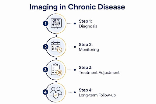
Diagnostic imaging is defined as the use of medical technologies to visualise the structure and function of the body for clinical decision-making. In chronic disease, imaging does far more than confirm a diagnosis. It guides treatment, tracks progression, and reveals changes that physical examination and blood tests simply cannot detect. Modalities like quantitative CT, MRI, and ultrasound have shifted the role of radiology in chronic conditions from a supporting tool to a central pillar of care. Understanding how imaging aids chronic disease assessment helps you ask better questions and make more informed decisions about your health.
What imaging techniques are used to diagnose and monitor chronic diseases?
Several core modalities form the foundation of diagnostic imaging for chronic diseases. Each has distinct strengths depending on the tissue, organ system, and clinical question involved.
- X-ray provides a fast, low-cost view of bones, lungs, and the chest. It remains the first-line tool for detecting structural changes in conditions like rheumatoid arthritis and chronic obstructive pulmonary disease (COPD).
- CT (computed tomography) produces detailed cross-sectional images and is particularly useful for lung disease, abdominal organs, and vascular structures. Quantitative CT goes further by measuring lung density and tissue composition numerically.
- MRI (magnetic resonance imaging) excels at soft tissue detail without radiation. It is the preferred modality for brain, spinal cord, joints, and bowel wall assessment.
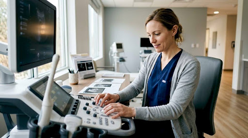
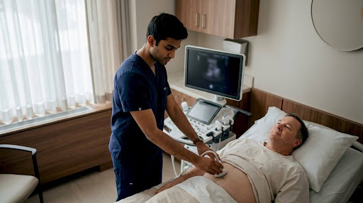
- Ultrasound is radiation-free, portable, and well-suited for real-time assessment of abdominal organs, blood vessels, and musculoskeletal structures. Vascular ultrasound identifies subclinical complications in conditions like diabetes and peripheral artery disease.
- PET (positron emission tomography) detects metabolic activity at the cellular level. It is used in cancer monitoring and increasingly in inflammatory disease.
Emerging tools like elastography measure tissue stiffness non-invasively, which is valuable for liver fibrosis staging. Advanced imaging modalities now monitor metabolic, functional, and molecular disease signatures, transitioning from anatomical detection to precision medicine. AI integration is accelerating image analysis and pattern recognition across all these modalities.
Pro Tip: Modality selection always balances diagnostic yield against safety. Radiation-free options like MRI and ultrasound are preferred for younger patients or those requiring frequent monitoring.
How does imaging contribute to managing COPD, diabetes, and IBD?
Imaging's contribution varies significantly across chronic conditions, but the common thread is that it reveals what clinical tests alone cannot.
COPD
COPD affects over 392 million people globally, with 3.3 million deaths annually. That scale demands precise monitoring tools. Spirometry measures airflow limitation, but quantitative CT goes further by measuring emphysema distribution, air trapping, and airway wall thickness. These metrics detect disease progression earlier and more accurately than lung function tests alone. Imaging is now core to COPD management, with standardised imaging scores integrated into clinical trials and routine care.
Diabetes
In diabetes, imaging detects organ damage before symptoms appear. Early imaging identifies hepatic steatosis (fat in the liver), pancreatic fat infiltration, and subclinical cardiovascular changes, enabling earlier risk stratification and treatment adjustment. This is clinically significant because organ damage in diabetes is often silent for years. Radiologists must also recognise that diabetic imaging findings can mimic malignancy or infection, making clinical context essential to avoid unnecessary procedures.
Inflammatory bowel disease
MRI and intestinal ultrasound now assess "transmural healing" in inflammatory bowel disease (IBD). Transmural healing refers to the resolution of inflammation across the full thickness of the bowel wall, a depth that endoscopy cannot reach. This imaging endpoint is now used to guide therapy decisions in Crohn's disease and ulcerative colitis.
Autoimmune and systemic inflammatory diseases
Ultrasonography, elastography, and MRI allow comprehensive evaluation of systemic complications in autoimmune diseases while reducing the need for invasive procedures like biopsies. This matters for patients who require long-term monitoring with minimal procedural burden.
| Chronic condition | Primary imaging modalities | Key metrics or findings |
|---|---|---|
| COPD | Quantitative CT, X-ray | Emphysema index, air trapping, airway wall thickness |
| Diabetes | Ultrasound, MRI, CT | Hepatic steatosis, pancreatic fat, vascular calcification |
| Inflammatory bowel disease | MRI, intestinal ultrasound | Transmural healing, bowel wall thickness |
| Autoimmune disease | MRI, ultrasound, elastography | Joint erosions, organ fibrosis, tissue stiffness |
| Chronic cardiovascular disease | Cardiac MRI | Myocardial hypertrophy, fibrosis, remodelling |
Serial cardiac MRI monitors remodelling in chronic cardiovascular disease, tracking hypertrophy and fibrosis in response to therapy. That level of detail shapes treatment decisions in a way that an echocardiogram alone cannot.
What are the safety and practical considerations for repeated imaging?
Repeated imaging in chronic disease is often necessary, but it requires careful justification. The key principle guiding this is ALARA, which stands for As Low As Reasonably Achievable. The ALARA principle guides clinicians to minimise cumulative radiation exposure, particularly for younger patients who face decades of monitoring ahead.
Practical considerations for patients include:
- Radiation accumulation: CT and X-ray involve ionising radiation. Repeated scans over years add up, and the risk is proportional to cumulative dose.
- Radiation-free alternatives: MRI and ultrasound carry no radiation risk and are preferred wherever they provide equivalent diagnostic information.
- Scan frequency: Imaging intervals should match clinical need. More frequent scans are not always better and should be driven by changes in symptoms or treatment response.
- Access and cost: Scan availability, wait times, and out-of-pocket costs vary. Discussing options with your healthcare provider helps you plan realistically.
- Patient comfort: MRI scans can be lengthy and feel confining. Ultrasound is quick and well-tolerated. Knowing what to expect reduces anxiety.
You can find useful context on lifetime X-ray safety to understand how radiation exposure is assessed over time.
Pro Tip: Keep a personal record of your imaging history, including dates, modalities, and the body regions scanned. Share this with every new provider to avoid duplicate or unnecessary studies.
What emerging imaging trends are shaping chronic disease care?
The importance of imaging in management is growing as new technologies extend what clinicians can see and measure.
- Whole-body MRI provides a comprehensive view of systemic inflammation in one scan. Whole-body MRI identifies asymptomatic disease regions in rheumatology that localised scans routinely miss. This is particularly valuable in conditions like axial spondyloarthritis, where inflammation spans multiple spinal segments.
- Radiomics and AI extract quantitative features from images that the human eye cannot detect. These tools build predictive models for disease progression and treatment response, moving imaging closer to personalised medicine.
- Hybrid molecular imaging combines PET with CT or MRI to capture both anatomy and metabolic activity in a single session. This is already standard in oncology and is expanding into inflammatory disease.
- Elastography measures organ stiffness non-invasively. It is now widely used to stage liver fibrosis in chronic liver disease, replacing many diagnostic biopsies.
Diagnostic radiology's shift towards precision medicine enables clinicians to track metabolic, functional, and molecular changes for personalised treatment. The challenge is implementation. Many of these tools require specialised equipment, trained radiologists, and clear evidence of cost-effectiveness before they become standard care. Coordinating these appointments efficiently matters too. Tools like a shared medical agenda are helping clinics manage complex imaging workflows for patients with multiple chronic conditions.
Key takeaways
Imaging is the most direct and non-invasive method available for tracking chronic disease progression, guiding therapy, and detecting complications before they become symptomatic.
| Point | Details |
|---|---|
| Imaging goes beyond diagnosis | Quantitative CT and MRI track disease progression and guide treatment adjustments over time. |
| Each modality has a specific role | Matching the right modality to the clinical question improves accuracy and reduces unnecessary exposure. |
| ALARA guides radiation safety | Cumulative radiation from repeated CT and X-ray must be minimised, especially in younger patients. |
| Emerging tools enable precision care | Whole-body MRI, radiomics, and hybrid PET imaging are extending what chronic disease monitoring can detect. |
| Clinical context is non-negotiable | Imaging findings must always be interpreted alongside patient history to avoid misdiagnosis. |
Why I think we underestimate what imaging tells us
After years of working in diagnostic imaging, the pattern I see most often is this: patients arrive after months of inconclusive blood tests and clinical exams, and a single well-chosen scan changes everything. Not because the scan is magic, but because chronic disease hides in tissue changes that no blood panel captures.
What concerns me more than imaging overuse is imaging underuse in the right context. A patient with longstanding Crohn's disease who has never had an MRI of the bowel may be managed on outdated information. A person with type 2 diabetes who has never had a liver ultrasound may have significant hepatic steatosis that nobody has addressed.
The technology has moved faster than clinical habits. Quantitative CT metrics for COPD exist and are validated, yet many patients still receive only spirometry results. Whole-body MRI for rheumatological disease is available, yet most patients with axial spondyloarthritis are still monitored with regional scans.
The future I am genuinely optimistic about is one where imaging findings are integrated into shared decision-making from the start. Not ordered reactively, but planned as part of a monitoring strategy. That shift requires both clinician education and patient engagement. You have a role in that too. Ask what imaging is appropriate for your condition, and ask how often it should be reviewed.
Cdncare's imaging services for chronic disease management
Cdncare operates across more than 20 clinic locations in Ottawa, with a team of over 150 radiologists and technologists dedicated to accurate, timely results. For patients managing chronic conditions, access to the right imaging at the right time makes a real difference. Cdncare offers ultrasound services that are radiation-free and well-suited for ongoing monitoring of abdominal organs, vascular structures, and soft tissue. Walk-in access is available through urgent care imaging for patients who need timely assessment without a long wait. Results are typically available within 24-48 hours, supporting faster conversations with your care team.
FAQ
What is the role of imaging in chronic disease?
Imaging provides non-invasive, detailed information about disease progression, organ changes, and treatment response that clinical exams and blood tests cannot deliver alone.
Which imaging modality is safest for long-term monitoring?
MRI and ultrasound involve no ionising radiation and are preferred for patients requiring frequent or long-term monitoring in chronic disease management.
How does quantitative CT differ from standard CT in COPD?
Quantitative CT measures lung density, emphysema distribution, and airway dimensions numerically, providing more sensitive progression data than standard visual CT interpretation or spirometry.
Can imaging detect diabetic complications before symptoms appear?
Yes. Imaging can identify hepatic steatosis, pancreatic fat infiltration, and subclinical cardiovascular changes in people with diabetes before any clinical symptoms develop.
What is transmural healing in inflammatory bowel disease?
Transmural healing refers to the resolution of inflammation across the full thickness of the bowel wall, an endpoint now assessed by MRI and intestinal ultrasound that endoscopy cannot reach.
Recommended
- Radiology in 2020 | Canadian Diagnostic Network
- Charlotte Imaging | Canadian Diagnostic Network
- The Importance of Vascular Ultrasound in Diagnosing Disease | Canadian Diagnostic Network
- Urgent Care Imaging | Ultrasound & X-Ray (Walk-In) | CDN
Your examination or doctor’s visit is 4 easy steps away