We now offer x-ray appointments at our Phenix, Charlotte, Rideau Valley, Nepean, Kanata, and Hunt Club clinics. Please check location availability before visiting.

Request an Appointment
Should you get an echocardiogram for a heart murmur?

An echocardiogram is the main test doctors use to find out whether a heart murmur reflects a problem with your heart’s valves or chambers. If your doctor heard a new murmur, or one that came with symptoms, an echo is almost always the next step. It is painless, non-invasive, and usually settles the question within days.
Most people who get one for this reason fall into a few groups:
- You have a newly detected murmur along with symptoms like shortness of breath or fatigue.
- Your doctor could not confirm on exam alone whether the murmur is harmless.
- You have a murmur that changed in character since a previous checkup.
Many echoes come back showing a normal heart with an innocent murmur. That result leads to reassurance, not urgent treatment.
Key Takeaways
An echocardiogram remains the most reliable first test for determining whether a heart murmur reflects normal blood flow or an underlying structural problem.
| Point | Details |
|---|---|
| Echo is the primary test | It is the standard first step for a symptomatic or unexplained murmur, per CorHealth Ontario guidance. |
| Most results are reassuring | Many murmurs turn out innocent, with a normal echo confirming no structural disease. |
| Test type depends on the case | TTE comes first; TEE, stress echo, and Doppler add detail when needed. |
| Symptoms change urgency | Chest pain, fainting, or severe breathlessness mean urgent care, not a routine booking. |
| Cdncare offers local access | Ottawa-area echocardiography with reports typically ready within 24 to 48 hours. |
This article is general information, not a substitute for advice from a qualified doctor. Consult a qualified healthcare professional about your own circumstances before acting on anything here.
Table of Contents
- What a heart murmur echocardiogram is and what it shows
- Why doctors order an echocardiogram for a murmur
- Types of echocardiogram tests and when each is used
- What happens during the test and how to prepare
- How echocardiogram results are interpreted and what happens next
- When to seek urgent care instead of a routine echocardiogram
- Limitations of echocardiography for murmurs
- Differential diagnosis: matching echocardiogram findings to murmur causes
- Is an echocardiogram safe? Risks and complications
- Alternative tests if an echocardiogram doesn’t give a clear answer
- How Cdncare supports your heart murmur workup
- Sources
- FAQ
What a heart murmur echocardiogram is and what it shows
An echocardiogram uses ultrasound, not radiation, to build live images of your heart as it beats. A sonographer moves a small transducer across your chest, and a cardiologist or radiologist later reviews the images to write the report. This combination, real-time motion plus expert interpretation, is what makes echo so useful for murmurs: a stethoscope tells your doctor that something sounds different, but the echo shows why.
The scan looks at:
- How your valves open and close, and whether blood leaks backward (regurgitation) or gets squeezed through a narrowed opening (stenosis).
- The size of your heart’s chambers and how forcefully they pump, expressed as ejection fraction.
- Blood flow patterns using Doppler ultrasound, which turns flow into colour and sound the reviewing physician can measure.
That level of detail separates an innocent murmur, common and harmless, from one caused by real structural change.
Why doctors order an echocardiogram for a murmur
Doctors do not send every murmur for imaging. The decision usually comes down to a few clinical signals: whether you have symptoms, whether the murmur is new, and whether the physical exam alone can rule out structural disease.
CorHealth Ontario’s echo standards treat echocardiography as the standard initial evaluation for a newly detected murmur in a symptomatic patient, and it applies just as strongly for an asymptomatic patient when the exam cannot confidently rule out structural disease. That second scenario surprises a lot of people. You can feel completely fine and still get referred for an echo, simply because certain murmurs cannot be safely dismissed on sound alone.
A murmur’s timing in the cardiac cycle, its grade on a standard loudness scale, and whether it radiates to your neck or back all shape how urgently your doctor wants imaging. A soft, early systolic murmur in a healthy teenager reads very differently than a loud, late murmur that radiates in an older adult.
A substantial number of children will have an innocent murmur detected at some point, which is one reason clinicians lean on these specific features, timing, grade, radiation, rather than treating every murmur the same way.
Types of echocardiogram tests and when each is used
Not every murmur needs the same test. Cardiologists choose among a few echo formats depending on what the initial exam suggests and what earlier imaging shows.
- Transthoracic echocardiogram (TTE) is the standard first test for almost everyone. It is done through the chest wall, gives a clear enough picture for most murmurs, and is where nearly every workup starts.
- Transesophageal echocardiogram (TEE) produces sharper images when a TTE is limited, which happens more often in patients with obesity, significant lung disease, or a prosthetic valve. The NHS’s overview of echo types explains why: sound waves travelling through the esophagus have a much shorter path to the heart than waves travelling through the chest wall and ribs.
- Stress or exercise echocardiogram reproduces murmurs or symptoms that only show up during exertion, and it doubles as a check for reduced blood flow to the heart muscle.
- Doppler and 3D echocardiography are add-on techniques layered onto TTE or TEE to measure exactly how much blood is leaking through a valve or how narrow an opening has become, information that shapes whether you’re monitored or referred for intervention.
What happens during the test and how to prepare
A standard TTE takes about 30 to 40 minutes, and there is no needle, no radiation, and generally no discomfort. You will lie on an exam table while a sonographer applies gel to your chest and moves the transducer to capture views from several angles. Expect to be asked to shift onto your left side or hold your breath briefly. Neither means anything is wrong; both simply give the sonographer a clearer acoustic window.

TEE is a different experience. You’ll need to fast beforehand, and because sedation is involved, you cannot drive for at least 24 hours afterward. Arrange a ride home before your appointment, not after.
For a stress echo, wear comfortable clothing and shoes you can walk or cycle in. Bring your requisition and a current medication list either way.
- TTE: no prep, no driving restriction, back to your day right after.
- TEE: fasting required, sedation involved, arrange transport home.
- Stress echo: comfortable footwear, expect to walk or cycle during imaging.
Pro Tip: Write down when the murmur was first noticed, along with any symptoms, even minor ones like occasional dizziness, and bring your prior cardiac records. It gives the interpreting physician context that speeds up the read.
How echocardiogram results are interpreted and what happens next
Most murmur workups end in one of two ways: a normal heart with an innocent murmur, or a specific structural finding that explains the sound. Many people fall into the first group. An echo that comes back clean simply confirms there’s no valve disease or structural issue driving the murmur, which usually means reassurance and no further testing.
When the report does show something, the cardiologist is reading a handful of specific measurements:
- Valve function, including whether a valve opens fully or closes tightly.
- Ejection fraction, the percentage of blood pumped out with each heartbeat.
- Regurgitation or stenosis severity, usually graded mild, moderate, or severe.
Depending on those numbers, next steps range widely: no action at all, a repeat echo in a year or two to watch for change, medication to manage symptoms, or referral to a cardiologist for a more detailed workup. Mild valve findings are often followed with periodic imaging rather than treated right away.
If your results land in a patient portal before your doctor has explained them, resist the urge to interpret the raw numbers yourself. Terms like “trace regurgitation” sound alarming out of context but are often clinically insignificant. Wait for the conversation with your clinician.
When to seek urgent care instead of a routine echocardiogram
Most murmurs are not emergencies, and most echo referrals can wait for a scheduled appointment. But some symptoms mean you should not wait at all.
- New or worsening chest pain.
- Sudden, severe shortness of breath, especially at rest.
- Fainting, or feeling like you’re about to faint.
- New swelling in your legs or abdomen, or other signs of heart failure.
If any of these show up alongside your murmur, go to an emergency department or call your clinician right away rather than waiting for a booked echo slot. A murmur alone, without these symptoms, rarely needs same-day attention. It’s the combination that changes the urgency, not the murmur by itself.
Limitations of echocardiography for murmurs
Echo is powerful, but it is not perfect, and knowing its blind spots helps set realistic expectations. Image quality depends heavily on body habitus and lung disease, and in patients with obesity or significant lung conditions, sound waves have a harder time reaching the heart cleanly. That’s precisely why TEE exists as a fallback: it bypasses the chest wall entirely by imaging through the esophagus.
Echo also has a harder time with certain conditions regardless of body type. Small holes in the heart’s walls, subtle valve thickening in early disease, and some congenital abnormalities can be missed or underestimated on a standard TTE, particularly if the sonographer isn’t specifically looking for them. Coronary artery disease, one possible contributor to certain murmur presentations, is not something a standard echo evaluates directly at all, since it images structure and flow rather than the coronary arteries themselves.
There’s also an operator and interpretation factor. A sonographer’s positioning skill and a physician’s experience reading borderline images both influence accuracy, which is part of why clinics with dedicated cardiac sonographers and radiologists who read echoes regularly tend to produce more consistent reports.
None of this means echo is unreliable. For the vast majority of murmurs, it answers the clinical question clearly. But if your symptoms and echo results don’t line up, or if your doctor still has concerns after a normal echo, that’s a sign for additional testing, not a sign the first test was wasted.
Differential diagnosis: matching echocardiogram findings to murmur causes
An echo doesn’t just confirm “murmur present.” It helps sort murmurs into distinct diagnostic categories, and the pattern of findings often points toward one specific cause.
A murmur caused by aortic stenosis typically shows a thickened, restricted valve with elevated blood flow velocity across it on Doppler. Mitral regurgitation shows up as backward-flowing blood on colour Doppler during the heart’s contraction phase, often with a specific pattern depending on which part of the valve is affected. Innocent murmurs, by contrast, show normal valve structure, normal chamber size, and no abnormal flow at all. The murmur exists, but the echo finds nothing wrong to explain it beyond normal blood flow turbulence.
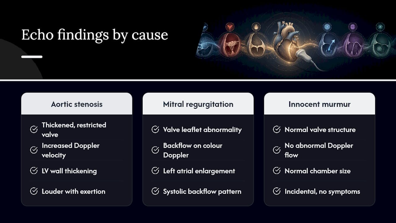
Chamber size and wall thickness add another layer. An enlarged left atrium alongside mitral valve changes suggests the murmur has been present long enough to affect heart structure over time. A thickened left ventricle paired with a murmur can point toward outflow obstruction rather than a leaking valve. These distinctions matter because treatment for a leaking valve looks nothing like treatment for a stiff, narrowed one.
This is also where your symptom history earns its place in the diagnostic picture. A murmur that gets louder with exertion, paired with specific Doppler findings, tells a different story than one found incidentally during an unrelated exam. The echo provides the anatomy and physiology; your clinical history helps the cardiologist decide which of several possible diagnoses actually fits.
Is an echocardiogram safe? Risks and complications
A standard transthoracic echocardiogram carries essentially no physical risk. It uses sound waves rather than radiation, involves no needles or dye, and the only real downside for most patients is mild discomfort from the pressure of the transducer against the chest, particularly if you have tender ribs or recent chest surgery. There is no recovery time and no restriction on driving or activity afterward.

TEE carries more to consider, because it is a mildly invasive procedure. Risks include a sore throat afterward, a small chance of reaction to the sedative, and rare but serious complications like injury to the esophagus, which occurs in a very small fraction of cases. This is why fasting beforehand and having someone drive you home matter: sedation affects reflexes and judgment for hours after the procedure ends.
Stress echocardiography adds the physical demands of exercise (or a medication that simulates it) on top of imaging. For most patients this is safe and well-monitored, but anyone with unstable chest pain or certain heart rhythm problems may need a different test instead, which your cardiologist will screen for before scheduling.
Across all versions of the test, the actual risk profile is low compared to many other cardiac investigations, particularly those involving catheters or radiation. That is part of why echo remains the default first step rather than something reserved for complicated cases.
Alternative tests if an echocardiogram doesn’t give a clear answer
Occasionally an echo comes back inconclusive, whether from limited image quality, a borderline finding, or symptoms that don’t match what the scan shows. When that happens, a few other tools come into play.
Cardiac MRI gives extremely detailed images of heart muscle and structure without the acoustic limitations that affect ultrasound, making it useful when body habitus or lung disease clouded the echo. A cardiac CT scan can map coronary arteries and calcium buildup, useful when chest pain accompanies the murmur and coronary disease needs to be ruled out. An electrocardiogram, while it cannot see structure at all, often runs alongside echo to check for rhythm problems or signs of strain on the heart, and understanding what an ECG actually measures helps explain why the two tests are frequently ordered together rather than as substitutes for one another.
For murmurs tied to exertion that a resting or even stress echo didn’t fully capture, a cardiac catheterization may eventually be considered, though this is invasive and generally reserved for cases where non-invasive imaging left a real diagnostic gap. Your cardiologist chooses among these based on exactly what the echo couldn’t answer, not as a routine next step for every inconclusive result.
A clinician’s perspective on what patients actually need to hear
Many patients come in unsettled by technical language in a portal report before anyone has explained what it means. Bring your prior records and a written symptom list; it shortens the appointment. If your murmur came with new symptoms, ask directly about expedited scheduling. Waiting weeks for clarity on a symptomatic murmur helps no one.
How Cdncare supports your heart murmur workup
Cdncare runs one of the largest diagnostic imaging networks in the Ottawa area, with sonographers and radiologists who read cardiac studies regularly rather than occasionally. That matters for a test where image quality and interpretation experience both affect accuracy. Our echocardiography services cover standard TTE studies for murmur evaluation, with reports typically turned around within 24 to 48 hours so you and your doctor aren’t left waiting on an answer.
Bring your physician’s requisition, a list of current medications, and any prior cardiac records to your appointment. If your symptoms feel urgent rather than routine, our urgent care imaging locations offer walk-in access without a long wait for a scheduled slot. Once your doctor has ordered the test, book your echocardiogram at a clinic near you and get the clarity you need to move forward.
Sources
- CorHealth Ontario — echo standards
- NHS — echocardiogram overview
- MyHealth Alberta — aftercare guidance for echocardiogram
FAQ
Should you worry if you have a heart murmur?
Not automatically. Many murmurs, especially in children and healthy adults, are innocent and never require treatment, but a new or symptomatic murmur still deserves an echocardiogram to rule out a structural cause.
Should you get an echocardiogram if you have a heart murmur?
If the murmur is new, symptomatic, or cannot be confidently explained on physical exam alone, yes. It is the standard first imaging test for exactly that situation.
What is a worrisome heart murmur?
A murmur that is loud, occurs late in the heartbeat cycle, radiates to the neck or back, or comes with symptoms like chest pain, fainting, or breathlessness carries more clinical weight than a soft, early, isolated murmur.
What does a cardiologist do if you have a heart murmur?
A cardiologist typically orders an echocardiogram to check valve function and heart structure, then decides between reassurance, periodic monitoring, medication, or further intervention based on what the scan and your symptoms show together.
Recommended
Your examination or doctor’s visit is 4 easy steps away