We now offer x-ray appointments at our Phenix, Charlotte, Rideau Valley, Nepean, Kanata, and Hunt Club clinics. Please check location availability before visiting.

Request an Appointment
What is mammography? A Canadian guide for 2026

TL;DR:
- Mammography is a low-dose X-ray technique that helps detect breast cancer early before symptoms appear. It involves two types: screening for healthy individuals and diagnostic for symptomatic cases, with guidelines suggesting biennial screening for ages 40 to 74. The procedure is safe, quick, and most abnormal results do not confirm cancer, emphasizing the importance of informed decision-making.
Mammography is a low-dose X-ray imaging method designed to detect breast cancer early, often before any symptoms appear. It produces detailed pictures of breast tissue that a radiologist then reviews for signs of abnormality. Screening mammography targets people with no symptoms, while diagnostic mammography investigates a specific concern. Canadian guidelines recommend routine screening for individuals aged 40 to 74, and early detection through mammography is directly linked to better survival outcomes. Understanding what to expect from the process makes it far easier to decide when and how to proceed.
What is mammography and how does it work?
Mammography uses low doses of radiation to produce X-ray images of the breast. The radiation dose is low, comparable to natural background exposure you encounter in everyday life, which means the safety profile is well established. The benefit of catching a small, treatable cancer far outweighs the minimal radiation risk.
Two main types of mammograms are used in clinical practice today. Standard 2D digital mammography captures flat images of the breast from two angles. Digital tomosynthesis, often called 3D mammography, takes multiple images at different angles and reconstructs them into a layered view of the breast tissue. Tomosynthesis improves diagnostic accuracy, particularly in patients with dense breast tissue, because it reduces the overlap that can obscure small lesions on a flat image.
During the mammogram procedure, a technologist positions your breast on a flat plate. A second plate then compresses the breast firmly for a few seconds while the image is captured. Compression is necessary because it spreads the tissue, reduces the radiation dose needed, and produces a sharper image. Most patients describe the sensation as pressure rather than pain, and each compression lasts only a few seconds.
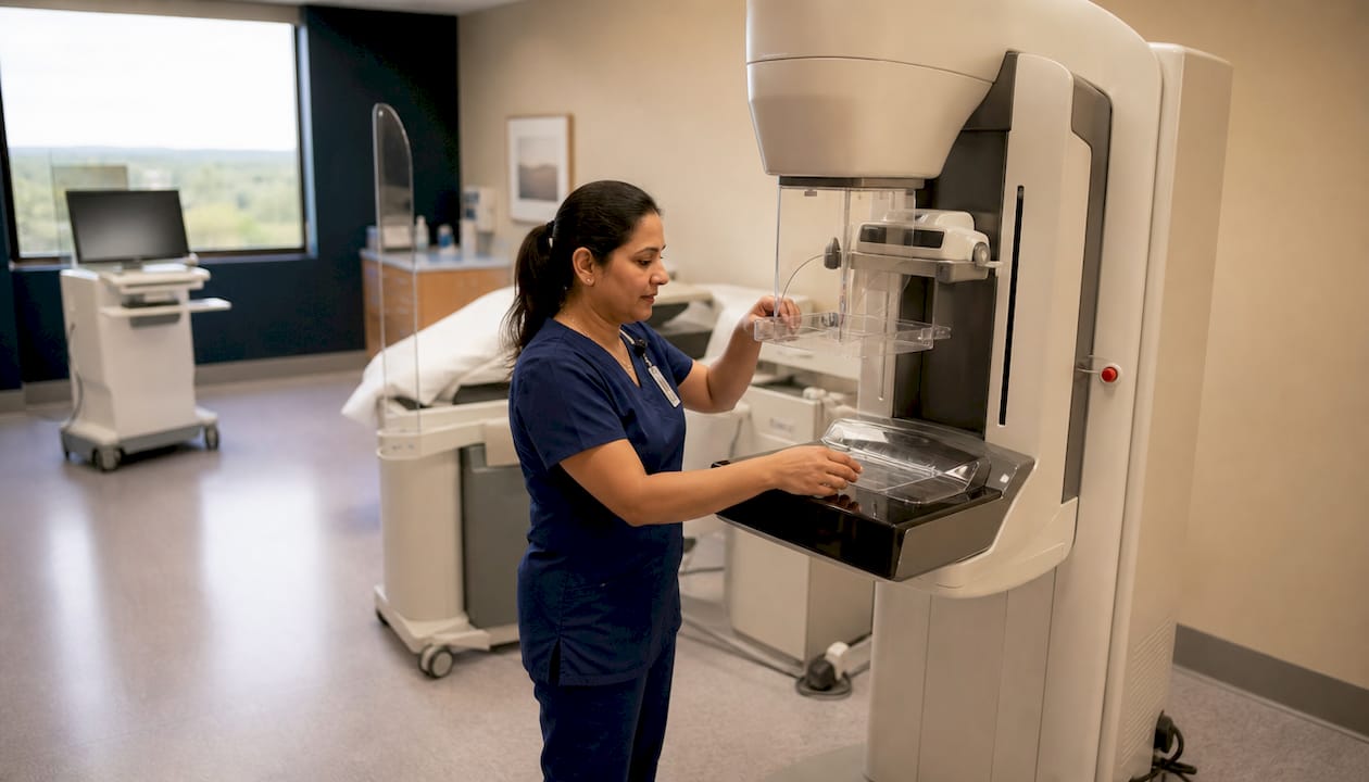
Pro Tip: If you are breastfeeding, pump or nurse immediately before your appointment. Reducing breast fullness beforehand improves both your comfort during compression and the clarity of the images.
Who should get mammography screening and when?
Canadian guidelines recommend biennial mammography for individuals aged 40 to 74 without elevated risk factors. The Ontario Breast Screening Programme (OBSP), one of the largest population-based screening networks in the country, follows this framework. Screening is open to women, as well as Two-Spirit, trans, and nonbinary individuals who have breast tissue, regardless of gender identity.
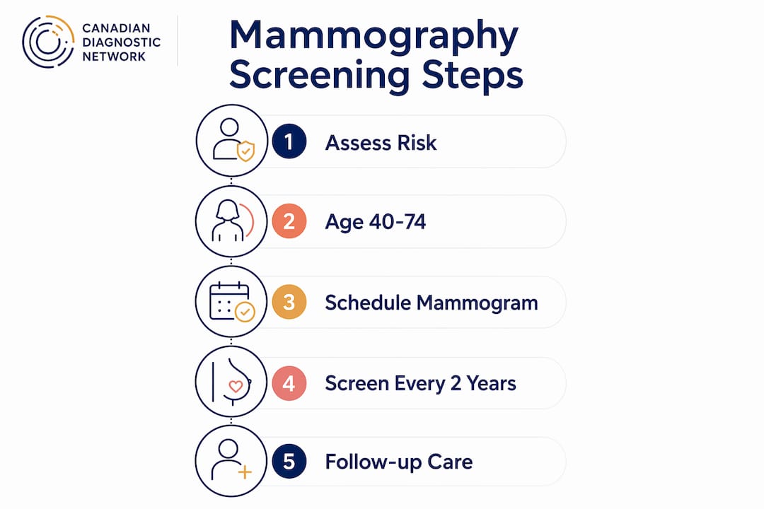
Your personal and family history can shift the recommended timing. A first-degree relative diagnosed with breast cancer, a known BRCA1 or BRCA2 gene mutation, or a history of chest radiation before age 30 all place you in a higher-risk category. In those cases, annual screening or supplemental imaging such as MRI may be recommended earlier than age 40. BRCA gene testing can clarify your risk level if your family history raises concern.
Shared decision-making between you and your provider is the foundation of good screening practice. Your values, your risk tolerance, and your personal health history all shape the right approach for you. No single schedule fits everyone.
Key criteria that influence your screening eligibility and timing:
- Age 40 to 74 with no elevated risk factors: biennial screening is the standard recommendation.
- Family history of breast or ovarian cancer in a first-degree relative: discuss earlier or more frequent screening with your provider.
- Known genetic mutation (BRCA1, BRCA2, or equivalent): annual screening and supplemental MRI are typically recommended.
- Previous chest radiation before age 30: earlier and more frequent screening applies.
- Dense breast tissue identified on a prior mammogram: supplemental ultrasound or tomosynthesis may be considered.
If you are unsure where you fall, your family doctor or nurse practitioner can help you assess your risk and connect you with the right programme.
Benefits and limitations of mammography for breast cancer detection
Screening mammography is considered the gold standard for population-based breast cancer screening, with demonstrated mortality benefits. Detecting a tumour when it is small and localised gives you more treatment options and a significantly better prognosis. That is the core case for routine screening.
The limitations are real and worth understanding. False positives occur when a mammogram flags something that turns out not to be cancer. This leads to additional imaging or biopsy, which creates anxiety and inconvenience. Overdiagnosis is a separate concern: some slow-growing cancers detected by screening may never have caused harm during a person’s lifetime, yet they still receive treatment. Missed cancers also happen, particularly in people with very dense breast tissue where tumours can be harder to see on a 2D image.
Transparent communication about both the benefits and limitations of mammography is the foundation of patient-centred care. Patients who understand the full picture are better equipped to make decisions that reflect their own values and circumstances.
Balancing these factors is not simple. The evidence supports screening, but the right frequency and the role of supplemental imaging depend on your individual situation. That is why informed consent and open dialogue with your provider matter as much as the test itself.
What to expect during a mammogram and follow-up
A mammogram appointment typically takes 20 to 30 minutes from arrival to departure. The imaging itself lasts only a few minutes. You will be asked to undress from the waist up and will be given a gown. The technologist will explain each step before it happens and will adjust positioning to suit your body.
Here is what the process looks like from start to finish:
- Arrival and check-in: You confirm your health history and any recent breast changes.
- Positioning: The technologist positions each breast on the imaging plate, one at a time.
- Compression: The plate compresses the breast briefly while the image is captured. You will feel pressure.
- Image review: The technologist checks image quality before you leave.
- Results: A radiologist reads the images and sends a report to your referring provider, typically within a few days.
Most patients receive a normal result. About 1 in 10 patients with an abnormal result are ultimately diagnosed with cancer after follow-up. An abnormal result means further assessment is needed, not that cancer is confirmed. Follow-up may include additional mammogram views, a breast ultrasound, or a biopsy.
Pro Tip: Scheduling your appointment in the week after your period, when breast tissue is less tender, can reduce discomfort. If you have concerns about pain during the procedure, speak with the technologist before you begin. They can adjust positioning to improve your comfort.
Key takeaways
Mammography is the most evidence-supported tool for early breast cancer detection, and understanding the process reduces anxiety and improves screening uptake.
| Point | Details |
|---|---|
| Core purpose | Mammography uses low-dose X-rays to detect breast cancer before symptoms appear. |
| Two types available | Screening mammography targets healthy individuals; diagnostic mammography investigates symptoms or abnormalities. |
| Canadian age guidelines | Biennial screening is recommended for individuals aged 40–74 without elevated risk factors. |
| Results in context | Only about 1 in 10 abnormal results leads to a cancer diagnosis after follow-up testing. |
| Shared decision-making | Your personal risk, values, and health history should guide screening frequency and supplemental imaging choices. |
Mammography fits into a bigger picture
Patients often arrive at their first mammogram carrying a mix of anxiety and uncertainty. That is completely understandable. What I have noticed, both from working in diagnostic imaging and from conversations with patients over the years, is that the anxiety usually comes from not knowing what to expect, not from the procedure itself.
What concerns me more is the tendency to treat mammography as either the complete answer or as something to avoid because of its limitations. Neither position serves patients well. Mammography is a strong tool for population-level screening, but it works best when it is part of a broader conversation about your individual risk. A patient with dense breast tissue and a family history needs a different plan than someone with average risk and no prior findings. Treating everyone identically misses the point of modern screening.
The other thing worth saying plainly: an abnormal result is not a diagnosis. Many patients I have spoken with describe the days between an abnormal result and follow-up imaging as the most stressful part of the entire experience. Knowing in advance that most abnormal results do not lead to a cancer diagnosis genuinely helps people stay calm and follow through with the next steps.
The best outcome comes from an informed patient who has had an honest conversation with their provider, understands the benefits and the limitations, and shows up for their appointment.
— Felix
Cdncare mammography services in Ottawa
Cdncare operates one of the largest Ontario Breast Screening Programme networks in the region, with digital mammography and tomosynthesis available across more than 20 clinic locations in Ottawa. Results are typically available within 24 to 48 hours, and the team includes over 150 radiologists and technologists trained in breast imaging. Whether you are booking a routine screening or following up on a previous result, Cdncare offers patient-focused care with clear communication at every step. You can learn more about mammography services or find a location near you to book your appointment.
FAQ
What is the difference between screening and diagnostic mammography?
Screening mammography is for people with no symptoms and aims to detect cancer early. Diagnostic mammography is used when a symptom, lump, or abnormality has already been identified and needs further investigation.
How long does a mammogram take?
A mammogram appointment typically takes 20 to 30 minutes in total. The actual imaging portion lasts only a few minutes.
Are mammograms safe?
Yes. Mammograms use a low radiation dose that is comparable to natural background exposure, and the risk from radiation is minimal relative to the benefit of early cancer detection.
What happens if my mammogram result is abnormal?
An abnormal result means further assessment is needed, not that cancer is present. Only about 1 in 10 patients with an abnormal result receive a cancer diagnosis after follow-up testing.
At what age should I start getting mammograms in Canada?
Canadian guidelines recommend starting biennial screening at age 40 for individuals at average risk. Those with elevated risk factors, such as a family history or a genetic mutation, may benefit from starting earlier.
Recommended
Your examination or doctor’s visit is 4 easy steps away