We now offer x-ray appointments at our Phenix, Charlotte, Rideau Valley, Nepean, Kanata, and Hunt Club clinics. Please check location availability before visiting.

Request an Appointment
CT scan vs MRI: what's the difference and when does it matter?
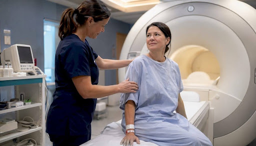
The core difference between CT and MRI scans comes down to the technology each uses and what that technology reveals. A CT scan uses X-rays to produce fast, detailed images of bones, organs, and blood vessels. An MRI uses strong magnetic fields and radio waves to create high-resolution images of soft tissues, with no ionizing radiation involved. Both are diagnostic imaging studies ordered by a licensed health professional, and both are interpreted by a radiologist.
Here is a quick summary of where each scan stands:
- CT scans produce images in seconds to minutes, making them the preferred choice in trauma and emergency settings
- MRI scans typically last from 15 minutes to over an hour but deliver superior detail for brain, spine, joint, and soft tissue conditions
- CT involves ionizing radiation; MRI does not, which matters for children and patients requiring repeated imaging
- MRI requires thorough metal screening before the scan; CT is generally safe for patients with metal implants
- Both may use contrast dye to improve image clarity, and both require a referral from a licensed health professional in Canada
What is an MRI and what does it show?
MRI uses powerful magnetic fields and radio waves to produce cross-sectional images of the body. No X-rays are involved, which means no ionizing radiation. This makes MRI particularly valuable when repeated imaging is needed, or when the patient is a child.
MRI excels at imaging soft tissues. Radiologists rely on it for:
- Brain and spinal cord conditions, including tumours, multiple sclerosis, and stroke assessment
- Joint and muscle injuries such as torn ligaments, rotator cuff tears, and cartilage damage
- Abdominal and pelvic organs, including the liver, kidneys, uterus, and prostate
- Cardiac structure and function
- Breast tissue assessment, often used alongside mammography
The level of soft tissue contrast MRI provides is genuinely difficult to match with other modalities. A ligament tear that would be invisible on a CT scan shows up clearly on MRI. For neurological assessments, MRI is often the first choice because it can distinguish between different types of brain tissue and detect subtle changes that CT would miss.
Pro Tip: If you have any metal implants, including pacemakers, surgical clips, or joint replacements, tell your technologist before your MRI appointment. Strong magnetic fields can move metallic objects and cause serious injury. Your care team will screen you carefully and determine whether MRI is safe for you.
What is a CT scan and what is it used for?
A CT scan combines multiple X-ray images, taken from different angles, to create detailed cross-sectional slices of the body. A computer processes those images to show bones, organs, and blood vessels in a way a standard X-ray cannot. CT scans make detailed images quickly, often in seconds to a few minutes.
CT is the workhorse of emergency and trauma imaging. Clinicians reach for it when speed matters most. Common uses include:
- Bone fractures and complex skeletal injuries
- Chest and lung conditions, including pulmonary embolism and pneumonia
- Abdominal emergencies such as appendicitis or internal bleeding
- Vascular conditions, including aneurysms
- Cancer staging and monitoring treatment response
- Head injuries where rapid assessment is critical
CT is also less restrictive than MRI for patients with metal hardware in their bodies. Someone with a hip replacement or surgical screws can typically undergo a CT scan without concern.
Pro Tip: CT scans involve ionizing radiation, which contributes to cumulative lifetime exposure. Your doctor weighs this carefully against the diagnostic benefit before ordering one. If you have had several CT scans in the past, mention this to your physician so they can factor it into the decision.
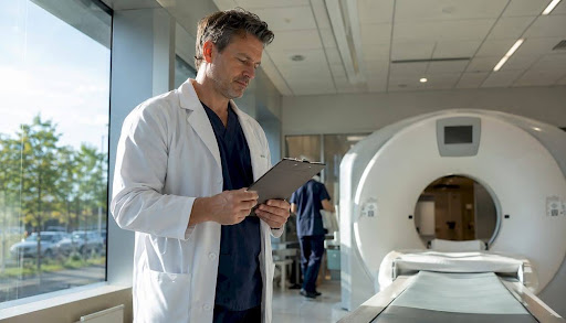
What to expect during an MRI scan
MRI appointments typically last from a moderate duration up to an extended period, depending on the area being scanned and the imaging protocol required. Brain and spine scans tend to run longer than a single joint study. You will lie on a table that slides into a narrow, tunnel-shaped machine.
The experience is manageable for most patients, but there are a few things to prepare for:
- Noise: The machine produces loud, rhythmic banging sounds throughout the scan. Ear protection is always provided.
- Stillness: You need to remain as still as possible. Movement blurs the images and may require the sequence to be repeated.
- Claustrophobia: The tunnel is narrow. If you feel anxious in enclosed spaces, let your care team know in advance. Some clinics can offer anti-anxiety medication to help you through the scan.
- Metal screening: Before entering the room, you will remove all metal objects and answer questions about implants, piercings, or any previous surgeries involving metal.
- Contrast dye: Some MRI scans require an injection of contrast material through an IV to improve image clarity. Your technologist will explain this beforehand.
Once the scan is complete, a radiologist interprets the images and sends a report to your referring physician, typically within a few days. MRI wait times in Canada tend to be longer than for CT, since MRI machines are in high demand and appointments take more time to complete.

What to expect during a CT scan
CT scans are fast. Most take only seconds to a few minutes from start to finish, which is one reason they are so well suited to emergency settings. The overall appointment, including preparation, is usually brief.
Here is what the experience looks like:
- You lie on a flat table that moves slowly through a large, doughnut-shaped scanner
- The machine is open on both sides, so claustrophobia is rarely an issue
- You may be asked to hold your breath briefly during certain chest or abdominal scans
- Contrast dye may be administered through an IV to highlight blood vessels or organs; if abdominal imaging is planned, you may need to fast beforehand
- The scan itself is quiet compared to an MRI, and most patients find it straightforward
Results are sent to your doctor within days. CT scans are more widely available across Canadian imaging centres than MRI, and wait times are generally shorter. In urgent situations, Canadian health systems use a triage level system from 1 to 4 to prioritise imaging resources, with emergencies seen immediately.
How do CT and MRI compare on risks, benefits, and preparation?
Both scans are safe when ordered appropriately, but they carry different considerations depending on your health history and what your doctor needs to see.
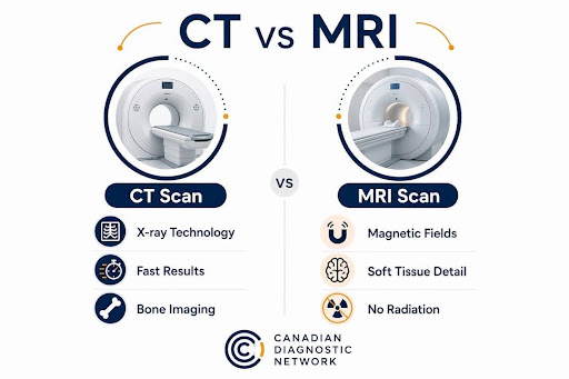
| CT scan | MRI scan | |
|---|---|---|
| Imaging technology | Multiple X-rays processed by computer | Magnetic fields and radio waves |
| Typical uses and strengths | Bone injuries, chest and lung, vascular, trauma, emergencies | Brain, spine, joints, soft tissues, organs, oncology staging |
| Risks and limitations | Ionizing radiation; not preferred for repeated use or in pregnancy | No radiation; metal implants may be a contraindication; poor calcium detection |
| Procedure experience | Fast, quiet, open scanner; low claustrophobia risk | Longer, loud, narrow tunnel; claustrophobia possible |
| Preparation requirements | Fasting if abdomen scanned; contrast dye possible | Metal screening required; contrast dye possible; anti-anxiety medication available |
One distinction worth noting: MRI does not detect calcium well, which matters in certain brain tumour assessments where calcification is a diagnostic clue. CT handles that clearly. Conversely, MRI provides soft tissue contrast that CT simply cannot match, which is why neurologists and orthopaedic specialists rely on it so heavily.
How do doctors decide between a CT scan and an MRI?
The choice between CT and MRI is rarely arbitrary. Physicians weigh several factors at once, and the decision often comes down to a combination of clinical urgency, the body part involved, and the patient's individual circumstances.
- Speed and urgency: In trauma or emergency situations, CT is preferred because it produces usable images within minutes. A patient with a suspected brain bleed or internal injury cannot wait an hour for an MRI.
- What needs to be seen: Soft tissue detail favours MRI. Bone, lung, and vascular structures favour CT. A suspected torn ACL calls for MRI; a suspected rib fracture calls for CT.
- Radiation considerations: When CT and MRI would provide comparable information, MRI is often preferred to avoid radiation exposure, particularly for children or patients who require ongoing monitoring.
- Metal implants: Patients with certain pacemakers, cochlear implants, or older surgical clips may not be eligible for MRI. CT is the safer alternative in those cases.
- Claustrophobia and patient tolerance: A patient who cannot tolerate the MRI tunnel may need a CT instead, or may require sedation before proceeding with MRI.
- Availability and wait times: In Canada, CT is more accessible. For non-urgent soft tissue assessments, a physician may order MRI knowing the patient will wait longer for the appointment.
For neurological conditions, the Canadian Medical Imaging Inventory notes that MRI is used across neurology, cardiology, oncology, and orthopaedics, while CT serves neurology, emergency trauma care, and oncology as well. The overlap is real, and the right choice depends on the specific clinical question being answered. If you are curious about how neurological imaging decisions are made, resources like the neurology CCRN study guide from Zero Deficit offer useful clinical context.
CT and MRI services available through Cdncare
Cdncare, the Canadian Diagnostic Network, offers both CT and MRI imaging across more than 20 clinic locations in the Ottawa area. With over 30 years of experience and a team of over 150 radiologists and technologists, CDN is positioned to support a wide range of diagnostic needs, from routine referrals to time-sensitive assessments.
What patients can expect from CDN's imaging services:
- Advanced MRI technology: CDN operates 1.5T and 3T MRI scanners, with 3T providing higher resolution for complex neurological and musculoskeletal cases
- High-resolution CT: 128-slice CT scanners deliver detailed cross-sectional images with fast acquisition times
- Rapid results: Reports are typically delivered to referring physicians within 24 to 48 hours
- Ontario Breast Screening Programme: CDN operates one of the largest OBSP networks in Ontario, integrating MRI and other modalities into breast cancer detection pathways
- Accessible, compassionate care: Patients are guided through preparation, screening, and the scan itself by experienced technologists who communicate clearly at every step
If you need imaging quickly, CDN's urgent care imaging service provides walk-in access to ultrasound and X-ray, with referrals to CT and MRI when clinically indicated. For patients in Ottawa, the Charlotte Imaging location offers comprehensive diagnostic services in a patient-focused setting.
Key takeaways
CT and MRI serve different diagnostic purposes: CT is faster and better for bones and emergencies, while MRI provides superior soft tissue detail without radiation exposure.
| Point | Details |
|---|---|
| Technology differs fundamentally | CT uses X-rays; MRI uses magnetic fields and radio waves with no ionizing radiation. |
| Speed favours CT | CT scans take seconds to minutes; MRI appointments typically last 15 minutes to over an hour. |
| Soft tissue detail favours MRI | Brain, spine, joints, and organs are imaged with higher contrast on MRI than CT. |
| Metal implants affect eligibility | Strong magnetic fields make MRI unsafe for some implants; CT is generally safe for patients with metal hardware. |
| Canadian wait times vary | MRI appointments tend to take longer to access than CT due to demand and machine availability. |
FAQ
Which is better, a CT scan or an MRI?
Neither is universally better. CT is faster and preferred for bone injuries, chest conditions, and emergencies, while MRI provides superior soft tissue detail for brain, spine, and joint assessments. Your doctor chooses based on what needs to be seen and how quickly.
What does an MRI show that a CT scan does not?
MRI reveals fine detail in soft tissues, including ligaments, tendons, spinal cord structures, and brain matter, with a level of contrast that CT cannot match. It is also the preferred tool for detecting subtle neurological changes and certain tumours in soft tissue.
Why would a doctor order a CT scan instead of an MRI?
CT is ordered when speed is critical, such as in trauma or suspected internal bleeding, or when the clinical question involves bones, lungs, or vascular structures. It is also chosen when a patient has metal implants that make MRI unsafe.
What are two major disadvantages of MRI scans?
MRI scans take significantly longer than CT scans, which limits their use in emergencies, and the strong magnetic field makes them unsafe for patients with certain metal implants such as older pacemakers or surgical clips. Claustrophobia can also be a barrier for patients who struggle with the narrow tunnel.
Recommended
Your examination or doctor’s visit is 4 easy steps away