We now offer x-ray appointments at our Phenix, Charlotte, Rideau Valley, Nepean, Kanata, and Hunt Club clinics. Please check location availability before visiting.

Request an Appointment
Examples of x-ray uses in medicine: a practical guide
TL;DR:
- X-ray imaging uses ionizing radiation to produce images that help diagnose injuries, diseases, and anatomical changes. It is a versatile tool in healthcare, with common applications including fracture detection, lung screening, dental diagnostics, and breast cancer screening. Modern low-dose digital technology ensures safety, while AI-assisted analysis promises faster, more accurate results.
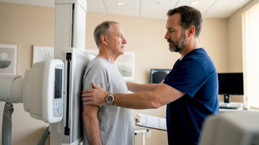
X-ray imaging is defined as a diagnostic technique that uses ionizing radiation to produce images of internal body structures, enabling clinicians to detect injury, disease, and anatomical changes without surgery. The examples of x-ray uses in medicine span from broken bones to breast cancer screening, making it one of the most versatile tools in healthcare. Medical radiation exposures from diagnostic sources account for about 47% of all radiation doses Canadians receive annually. That figure reflects just how central X-ray imaging has become to everyday clinical care. Most appointments take 10 to 15 minutes and are completely painless, which makes X-rays accessible for patients of all ages.
Bone fracture detection and skeletal assessment
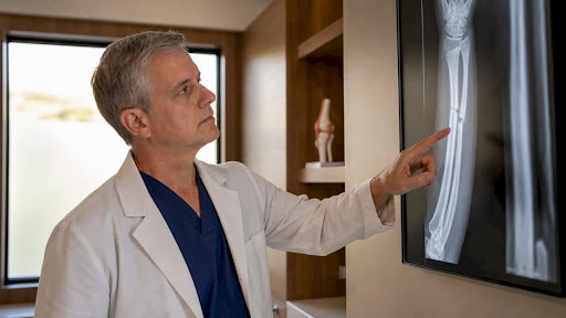
Bone and joint imaging represents the most common category of X-ray applications in healthcare. When you come in after a fall or injury, an X-ray gives your physician a clear picture of whether a bone is broken, cracked, or displaced. Radiologists also use skeletal X-rays to assess how well a fracture is healing over time, which guides decisions about when to remove a cast or modify a treatment plan.
X-rays are equally useful for detecting degenerative joint conditions like osteoarthritis, where the space between joints narrows visibly on film. Spine and pelvis imaging can reveal alignment problems, compression fractures, or signs of disc degeneration. Upper and lower extremity X-rays, covering everything from wrists to ankles, are among the most frequently requested studies in outpatient clinics.
- Fracture identification and monitoring of healing progress
- Arthritis staging and joint space assessment
- Spinal alignment and compression fracture detection
- Upper extremity studies: wrist, elbow, shoulder
- Lower extremity studies: ankle, knee, hip
Bone mineral density imaging is a specialised X-ray application that measures calcium levels in bone to assess osteoporosis risk and predict fracture likelihood. This scan provides quantitative data that is critical for managing long-term bone health.
Pro Tip: If you are being monitored for a healing fracture, bring any previous X-ray images to your follow-up appointment. Comparing images over time gives your radiologist the clearest picture of your progress.
Chest and lung imaging for respiratory conditions
The chest radiograph accounts for up to 20% of all imaging requested globally, making it the single most common radiological investigation in the world. That prevalence reflects its clinical value: a single chest X-ray can reveal pneumonia, lung tumours, fluid around the lungs, an enlarged heart, and signs of active tuberculosis.
Pneumonia appears on a chest X-ray as an area of increased density, often described as a "consolidation." Lung tumours may show up as rounded masses or irregular shadows. Tuberculosis produces characteristic patterns in the upper lobes of the lungs, which is why chest X-rays are a standard screening tool in public health settings.
- Pneumonia and lower respiratory tract infections
- Lung tumours and masses
- Pleural effusion (fluid around the lungs)
- Heart size and shape assessment
- Active tuberculosis identification
Chest X-rays for Canadian immigration applicants are a routine screening step for tuberculosis and do not indicate suspicion of disease. An abnormal finding does not automatically result in application refusal but may require follow-up testing.
Chest X-rays are one of the most informative and cost-effective diagnostic tools available. A single image can simultaneously assess the lungs, heart, ribs, and major blood vessels, providing a broad clinical overview in minutes.
Dental X-rays and oral health diagnostics
Dental radiography is one of the clearest examples of x-ray technology applied outside of hospital settings. Dentists use X-rays to detect cavities between teeth that are invisible to the naked eye, assess bone loss around tooth roots, and identify impacted teeth before they cause pain or infection.
Periapical X-rays show the full length of individual teeth, including the root and surrounding bone. Panoramic X-rays capture the entire jaw in a single image, which is useful for planning orthodontic treatment or evaluating wisdom teeth. Digital dental X-rays use significantly lower radiation doses than older film-based systems, making routine dental imaging safer than ever.
Mammography for breast cancer screening
Mammography uses low-dose X-rays to image breast tissue and detect cancer at its earliest, most treatable stage. The Ontario Breast Screening Program (OBSP), in which Cdncare operates one of the largest networks in the province, uses mammography as its primary screening tool for eligible patients.
Early detection through mammography significantly improves treatment outcomes. Cdncare's mammography services are available across multiple Ottawa locations, with results typically returned within 24 to 48 hours. If a mammogram identifies an area of concern, follow-up imaging such as ultrasound or a targeted biopsy may be recommended.
- Screening mammography for patients with average risk, typically starting at age 50
- Diagnostic mammography for patients with a lump, pain, or other breast symptom
- High-risk screening for patients with a family history or BRCA gene mutation
Fluoroscopy: real-time X-ray imaging
Fluoroscopy provides real-time moving X-ray images, which makes it fundamentally different from a standard static X-ray. Clinicians use it to watch physiological processes as they happen, such as how a patient swallows, how contrast material moves through the gastrointestinal tract, or how a joint moves under load.
Fluoroscopy is also used to guide interventional procedures. A radiologist can use live imaging to position a catheter, drain a fluid collection, or place a stent with precision. This real-time guidance reduces the risk of complications and improves procedural accuracy.
Bone density scanning for osteoporosis management
Bone mineral density (BMD) scanning is a low-radiation X-ray technique that measures calcium density in bone. It is the standard method for diagnosing osteoporosis and estimating fracture risk in patients over 50, post-menopausal women, and anyone on long-term corticosteroid therapy.
The scan is quick, non-invasive, and produces a score that your physician uses to guide treatment decisions. If you are curious about what happens during a bone density scan, the procedure involves lying still on a table while a low-dose X-ray arm passes over your body. Results are available to your physician within 24 to 48 hours at Cdncare.
Safety considerations and patient preparation
Radiation doses from diagnostic X-rays are low and regulated. Modern low-dose digital imaging protocols prioritise image quality and clinical necessity, and the minimal radiation exposure is medically justified by the diagnostic information gained. Routine lead shielding has been discontinued at major Canadian centres because updated equipment and scientific evidence show it is no longer necessary for patient safety.
Key preparation points for your appointment:
- Wear two-piece clothing without metal fasteners to avoid delays
- Remove jewellery, piercings, or accessories near the area being imaged
- Bring your physician's requisition, as clinics require a referral to perform non-emergency X-rays
- Inform the technologist if you are pregnant or may be pregnant
Pro Tip: Wearing clothing without metal fasteners to your X-ray appointment can meaningfully shorten your visit. Metal buttons, underwire, and zippers often require you to change into a gown, which adds time.
X-rays in treatment planning and ongoing monitoring
X-rays contribute to patient care well beyond the initial diagnosis. The table below outlines the most common ways X-ray imaging supports treatment decisions and long-term monitoring.
| Clinical situation | Role of X-ray imaging |
|---|---|
| Fracture management | Confirms alignment and monitors healing at follow-up visits |
| Arthritis progression | Tracks joint space narrowing over months or years |
| Osteoporosis treatment | BMD scans measure response to medication over time |
| Cancer diagnosis | Identifies tumours, metastases, and treatment response |
| Interventional procedures | Guides catheter placement, drainage, and stent insertion |
The relationship between imaging findings and clinical decisions is direct. A radiologist's report describing fracture alignment, for example, tells a surgeon whether a patient needs an operation or can heal with a cast. That specificity is what makes X-ray usage in diagnostics so valuable across specialties.
Key takeaways
X-ray imaging covers a wider range of clinical applications than most patients realise, from fracture detection to cancer screening, and each use is supported by clear safety protocols and modern low-dose technology.
| Point | Details |
|---|---|
| Bone and joint imaging | X-rays detect fractures, arthritis, and alignment problems across the full skeleton. |
| Chest X-ray prevalence | Chest radiographs account for up to 20% of all imaging globally and screen for infections, tumours, and TB. |
| Specialised applications | Mammography, dental radiography, and fluoroscopy extend X-ray use into cancer screening and real-time imaging. |
| Safety standards | Modern digital X-rays use low doses; lead shielding is no longer routine in Canadian clinics. |
| Treatment monitoring | X-rays guide ongoing care for fractures, osteoporosis, arthritis, and cancer, not just initial diagnosis. |
What I have learned watching X-ray technology evolve
The shift from film to digital radiography changed the patient experience in ways that are easy to underestimate. Images are available within seconds rather than minutes, doses are lower, and the image quality is sharper. Patients who were anxious about radiation exposure a decade ago are often surprised to learn how much the technology has improved.
What I find most underappreciated is how much a single X-ray communicates. Patients sometimes arrive expecting a definitive answer to a complex problem, and it is worth being honest: X-rays show structure, not function. They tell you a bone is broken, not why it broke. They show a shadow in the lung, not what caused it. That distinction matters, because it sets realistic expectations and explains why follow-up tests are sometimes needed.
The next shift I anticipate is AI-assisted X-ray analysis, where algorithms flag subtle findings that a human eye might miss on a busy day. That does not replace the radiologist. It gives them a second set of eyes on every image. For patients, that means faster, more consistent results. That is a meaningful step forward.
X-ray services at Cdncare: walk-in and urgent care imaging
Cdncare offers walk-in urgent care imaging across more than 20 Ottawa locations, with X-ray results typically returned within 24 to 48 hours. Whether you need a chest X-ray, a bone study, or a bone density scan, Cdncare's team of over 150 radiologists and technologists is equipped to handle a wide range of diagnostic X-ray exams. No long waits, no complicated booking process. You bring your physician's requisition, and the clinic takes care of the rest. Cdncare also provides mammography services through the Ontario Breast Screening Program, making it a single destination for many of the most common X-ray applications in Canadian healthcare.
FAQ
What are the most common examples of x-ray uses in medicine?
The most common uses include chest imaging for pneumonia and tuberculosis, bone fracture detection, dental cavity assessment, mammography for breast cancer screening, and bone density scanning for osteoporosis. Each application uses the same basic technology but targets different body systems and clinical questions.
Are X-rays safe for routine diagnostic use?
Yes. Modern digital X-rays deliver low radiation doses that are medically justified by the diagnostic value they provide. Canadian clinics follow regulated protocols, and routine lead shielding is no longer used because updated equipment makes it unnecessary.
Do I need a doctor's referral to get an X-ray in Canada?
Most Canadian diagnostic clinics require a physician's requisition to perform non-emergency X-rays. This is a legal and safety requirement, not a clinic policy. If you are unsure whether you need an X-ray, a guide on when to get an X-ray can help clarify the indications.
How long does an X-ray appointment take?
A standard diagnostic X-ray appointment typically takes between 10 and 15 minutes. The procedure itself is non-contact and painless, though preparation time may vary depending on the area being imaged and what you are wearing.
What is fluoroscopy and how does it differ from a standard X-ray?
Fluoroscopy produces real-time moving X-ray images rather than a single static picture. It is used to observe dynamic processes like swallowing, gastrointestinal movement, or joint motion, and to guide interventional procedures such as catheter placement.
Recommended
- How Many X-Rays Are Safe in a Lifetime?
- How to Tell If You Need an X-Ray | Canadian Diagnostic Network
- X-Ray | Canadian Diagnostic Network
- Finding the Difference Between an X-Ray and a Fluoroscopy
Your examination or doctor’s visit is 4 easy steps away