We now offer x-ray appointments at our Phenix, Charlotte, Rideau Valley, Nepean, Kanata, and Hunt Club clinics. Please check location availability before visiting.

Request an Appointment
Is an X-ray during pregnancy actually safe?
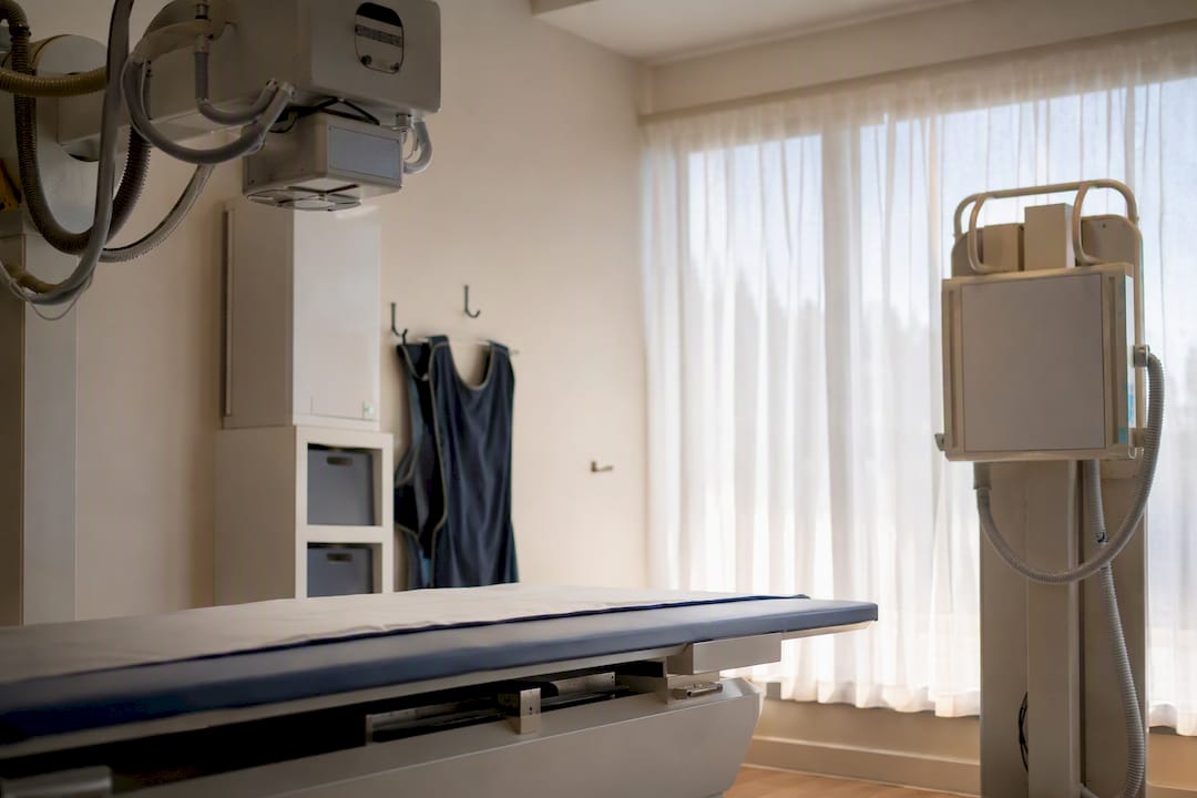
Most X-rays taken during pregnancy carry a very low risk to the fetus, and for exams like chest X-rays, dental X-rays, or images of your arms and legs, the exposure is small enough that it isn’t considered a meaningful safety concern. The exceptions worth knowing about are exams that aim directly at the pelvis or abdomen, or imaging that involves fluoroscopy over an extended period. If you’re pregnant or think you might be, the single most important thing you can do is tell your clinician or the imaging technologist before the exam happens.
- Routine exams (chest, dental, limbs) are unlikely to harm a fetus at typical diagnostic doses
- Pelvic X-rays, abdominal or pelvic CT, and repeated fluoroscopy are the main exceptions to flag
- Always disclose a known or suspected pregnancy before imaging, not after
- In an emergency, necessary imaging should still go ahead. Delaying it to avoid radiation can be riskier than the exposure itself
Key Takeaways
Most diagnostic X-rays deliver a fetal dose far below the 50 mGy threshold linked to developmental risk, making disclosure and exam-specific counselling more useful than blanket avoidance.
| Point | Details |
|---|---|
| Routine exams are low risk | Chest, dental, and extremity X-rays deliver under 0.01 mGy to the fetus, far below the 50 mGy concern threshold. |
| Pelvic and CT exams need more care | Pelvic X-rays run around 1.1 mGy, while abdominal or pelvic CT and fluoroscopy can go higher. |
| Disclose before, not just after | Telling your technologist beforehand allows shielding, collimation, and positioning adjustments in advance. |
| Emergencies shouldn’t wait | Necessary trauma or infection imaging protects the mother, which protects the fetus. |
| Cdncare documents and reviews every exam | Radiologists review dose and imaging details as part of standard workflow, with results typically in 24 to 48 hours. |
This article is general information, not a substitute for advice from a qualified doctor. Consult a qualified healthcare professional about your own circumstances before acting on anything here.
Table of Contents
- How x-ray during pregnancy exposure actually works
- Which x-rays carry the lowest risk, and which carry more
- Does timing in pregnancy change the risk?
- What about ultrasound and MRI instead of X-ray?
- What if you had an X-ray before you knew you were pregnant?
- How clinics reduce fetal exposure during necessary imaging
- When is an X-ray necessary despite pregnancy?
- How we approach imaging decisions for pregnant patients
- What conventional advice about X-rays in pregnancy gets wrong
- Get imaging support that takes pregnancy seriously
- Sources
- FAQ
How x-ray during pregnancy exposure actually works
An X-ray is a form of ionizing radiation, meaning it carries enough energy to knock electrons out of atoms and, in rare cases, damage DNA inside a cell. That sounds alarming out of context, but the dose from a diagnostic X-ray is tiny compared to what’s used in radiation therapy, and the body has repair mechanisms for the kind of minor cellular damage a routine scan might cause. During pregnancy, the developing fetus is more sensitive to radiation than an adult body simply because its cells are dividing and specializing rapidly, particularly in the first trimester.
Radiation dose to the fetus is measured in milligray (mGy), and having a reference point matters more than the number itself. A fetus absorbs roughly 0.5 mGy of natural background radiation over the course of an entire pregnancy just from cosmic rays and environmental sources. Health Canada and clinical guidelines generally treat 50 mGy as the conservative threshold below which there’s no established link to fetal malformation or pregnancy loss, and the doses from common diagnostic procedures sit well under that line.
Quick reference: dose context
- Background radiation over 40 weeks: about 0.5 mGy
- Conservative concern threshold: around 50 mGy
- Most single diagnostic X-rays: substantially below concerning exposure levels
The practical takeaway is that no single, appropriately performed diagnostic X-ray comes close to the dose levels associated with developmental harm. That gap is exactly why Health Canada’s public guidance states that correctly performed diagnostic procedures haven’t been shown to increase the risk of prenatal death, developmental damage, or impaired mental development at typical doses.
Which x-rays carry the lowest risk, and which carry more
The location of the exam matters far more than the fact that it’s an “X-ray” in general terms. When the X-ray beam isn’t aimed anywhere near your abdomen or pelvis, scatter radiation reaching the fetus is minimal, which is why these exams rarely require special precautions:
- Chest X-rays: fetal dose under 0.01 mGy, according to Health Canada’s estimates
- Dental X-rays: similarly under 0.01 mGy, with the beam directed at the jaw, far from the uterus
- Extremity X-rays (hands, feet, arms, legs): negligible fetal exposure since the beam never crosses the torso
A resource written for parents on pediatric foot X-rays makes a similar point about extremity imaging in general: when the beam is confined to a limb, scatter to distant organs is minimal regardless of who’s being imaged.
Exams that aim at or near the pelvis carry more dose, and it’s worth knowing the approximate ranges so you can ask informed questions:
- Pelvic X-ray: around 1.1 mGy in typical protocols
- Abdominal or pelvic CT: can range from several mGy up into the tens of mGy, depending on the protocol and number of passes
- Fluoroscopy (extended, real-time imaging): dose accumulates with time under the beam and can be the highest of the group
If you’re seeking counselling about a specific exam, give your care team the exact exam name, the body part imaged, and the date. That level of detail lets a radiologist or medical physicist calculate your actual fetal dose rather than guessing from a general category.
Does timing in pregnancy change the risk?
Gestational age changes how sensitive the fetus is to radiation, and it changes what kind of harm is theoretically possible. In the first two weeks after conception, the prevailing model is “all or none”: a high enough dose might end the pregnancy, but survivable exposure typically leaves no lasting effect, because cells at that stage aren’t yet differentiated into specific organs. From roughly week 3 through week 8, organogenesis is underway, and this window is the most sensitive period for structural malformation if the dose is high enough. Diagnostic X-rays, notably, stay far below the doses associated with that risk.
Later in pregnancy, the malformation risk drops substantially, but a different kind of risk becomes the relevant one: the fetus’s lifetime cancer risk rises roughly in proportion to dose, at any gestational age, though the absolute increase from a single diagnostic exam remains small.
- Weeks 0 to 2: all-or-none effect; low practical concern at diagnostic doses
- Weeks 3 to 8: peak sensitivity for structural effects, though still well under diagnostic-dose thresholds
- Second and third trimester: malformation risk falls, but any dose still carries a small cancer-risk increment
The practical implication is straightforward. If a pelvic X-ray or CT can reasonably wait until after delivery, many clinicians will suggest that. If it can’t wait, because you’re in pain, bleeding, or showing signs of a serious condition, it shouldn’t be delayed to avoid a dose that’s already well below the level of concern.
What about ultrasound and MRI instead of X-ray?
Ultrasound is the first choice for a wide range of obstetric and non-obstetric questions during pregnancy because it uses sound waves, not ionizing radiation, and it gives real-time images that work well for soft tissue, blood flow, and fetal assessment. Ultrasound covers everything from checking the gallbladder to evaluating suspected blood clots.
MRI is the next step up when ultrasound doesn’t give enough detail. MRI itself doesn’t use ionizing radiation, so it’s considered safe in the second and third trimesters when clinically indicated. Contrast agents used with MRI are a different matter: they cross the placenta, so radiologists use them sparingly and only when the diagnostic benefit clearly outweighs the theoretical risk.
- Ultrasound: no ionizing radiation, real-time, first-line for most pregnancy-related questions
- MRI: no ionizing radiation, used when ultrasound can’t answer the clinical question
- MRI contrast: crosses the placenta, used only when necessary
Pro Tip: If your doctor orders imaging and you’re pregnant, ask directly whether ultrasound or MRI could answer the same clinical question. Sometimes it can. Sometimes an X-ray or CT genuinely gives sharper, faster information, and that’s a reasonable outcome too.
What if you had an X-ray before you knew you were pregnant?
This comes up more than you’d think. Many patients come in after a trip to urgent care or a dental office, only to discover a few weeks later that they were already pregnant at the time. The anxiety that follows is understandable, but the math is usually reassuring.
- Contact the clinician or imaging centre that performed the exam. Give them the exam name, body part imaged, and the date.
- Ask them to estimate the fetal dose. For chest, dental, or extremity X-rays, this is typically a quick reassurance, since the dose is a small fraction of background exposure.
- If the exam involved the pelvis or abdomen, ask for a referral to radiology or maternal-fetal medicine for a more detailed dose calculation.
- Expect a conversation, not a diagnosis by phone. A radiologist may need imaging details from the technologist’s records to give you an accurate number.
Radiology departments generally keep detailed exposure records specifically so a pregnancy disclosed after the fact can be assessed properly. That documentation is what makes step one worth doing right away rather than searching for answers online.
How clinics reduce fetal exposure during necessary imaging
Radiology teams follow a principle called ALARA, “as low as reasonably achievable,” which shapes almost every decision made once a pregnant patient needs imaging. It starts with whether the exam can be postponed safely, or replaced with ultrasound or MRI, and continues into the technical details of the exam itself.
- Collimation: narrowing the X-ray beam so it covers only the area of clinical interest, reducing scatter
- Positioning: choosing posterior-anterior (PA) over anterior-posterior (AP) views where possible, which changes the beam’s path relative to the fetus
- Shielding: applying gonadal or abdominal shielding when it won’t interfere with the diagnostic image
- Dose documentation: recording exposure so a medical physicist can calculate fetal dose precisely if needed
Pro Tip: You don’t need to ask for shielding for a chest or dental X-ray. The fetal dose is already negligible for those exams, though many technologists will still offer a lead apron simply because it eases patient anxiety, not because the physics demands it.
When is an X-ray necessary despite pregnancy?
Some clinical situations don’t leave room for waiting. Trauma imaging after a fall or car accident, suspected pulmonary embolism, and severe infection are common examples where imaging can be genuinely lifesaving for the mother. The guiding clinical principle here is simple: appropriate maternal care is generally the best protection the fetus has.
- Trauma evaluation after a significant injury
- Suspected blood clot in the lungs
- Severe or worsening infection requiring imaging to locate the source
Expect your care team to talk you through the reasoning, including which technique they’re using and why, before proceeding. Urgent care imaging exists precisely for these time-sensitive situations, where the risk of delay outweighs the small radiation dose involved.
How we approach imaging decisions for pregnant patients
Cdncare has provided diagnostic imaging across the Ottawa area for over 30 years, with a team of more than 150 radiologists and technologists handling everything from routine X-rays to ultrasound and mammography. When a pregnant patient needs an X-ray, the process starts with your physician’s requisition, continues through the technologist confirming exam details with you, and ends with a radiologist reviewing the images, typically within 24 to 48 hours.
- Requisition reviewed against clinical need before scheduling
- Technologist confirms pregnancy status and exam details before proceeding
- Radiologist review and dose documentation completed as part of standard workflow
- Questions about a specific exam are always worth raising directly with the clinic, since decisions are made case by case
What conventional advice about X-rays in pregnancy gets wrong
The conventional wisdom tends to swing to extremes. Either an X-ray during pregnancy sounds catastrophic, or it gets waved off with a vague “it’s fine, don’t worry about it.” Neither version actually serves the reader. The dose numbers matter, and so does the exam type, and pretending otherwise does patients a disservice.

What gets underrated is how much the type of exam matters compared to the mere fact that radiation is involved. A chest X-ray and a pelvic CT aren’t remotely comparable in fetal dose, yet both get lumped into the same anxious mental category by a lot of patients. That’s backward. The conversation should start with “what body part, what modality, what date,” not “was radiation involved.”
What deserves more attention is timing of disclosure, not timing of the pregnancy itself. Telling a technologist before an exam gives the radiology team room to adjust technique. Finding out after the fact still allows for accurate dose reconstruction, but it removes the option to modify anything in advance. If there’s one habit worth building, it’s asking “should I mention I might be pregnant?” before every scan, not after.
— Felix
Get imaging support that takes pregnancy seriously
If you’re pregnant and need imaging, whether it’s a routine chest X-ray, an ultrasound to check something unrelated to the pregnancy, or an urgent scan after an injury, Cdncare gives you access to radiologists and technologists who factor pregnancy into every step of the process, from requisition through dose documentation. Unlike a walk-in clinic without dedicated imaging staff, Cdncare’s team reviews your exam details before you’re on the table, and turns around radiologist-reviewed results within 24 to 48 hours so you’re not left waiting on answers during an already anxious time. For non-obstetric questions where ultrasound can answer the clinical question without any radiation at all, book an ultrasound appointment directly. If your situation is urgent, urgent care imaging is available at multiple Ottawa-area locations without the wait of an emergency department.
Sources
- It’s Your Health: Diagnostic X-rays and Pregnancy (Health Canada publication)
- Non-obstetric diagnostic imaging in pregnancy (CMAJ)
FAQ
Is it safe to get an X-ray while pregnant?
Most X-rays, particularly chest, dental, and limb exams, deliver fetal doses far below levels associated with harm. Pelvic and abdominal imaging carries more dose and deserves a conversation with your care team first.
What happens if you get X-rays done while pregnant?
For low-dose exams like chest or dental X-rays, nothing measurable happens to the fetus at typical doses. For higher-dose exams like pelvic CT, your radiology team calculates the estimated fetal dose and discusses it with you.
What is the 10-day rule for radiology during pregnancy?
The “10-day rule” is an older radiology convention suggesting elective abdominal or pelvic X-rays be scheduled within the first 10 days after a menstrual period starts, when pregnancy is least likely. Many departments now rely on direct pregnancy screening questions instead, since it’s more reliable.
What are the do’s and don’ts during pregnancy regarding X-rays?
Do tell your clinician or technologist about a known or suspected pregnancy before any exam. Don’t delay medically necessary imaging out of fear. The risk of missing a serious diagnosis usually outweighs the small radiation dose involved.
Can you have an X-ray while pregnant if it’s an emergency?
Yes. Emergency imaging for trauma, suspected blood clots, or severe infection should proceed without delay, since untreated maternal conditions typically pose a greater risk to the fetus than the radiation dose itself.
Your examination or doctor’s visit is 4 easy steps away