We now offer x-ray appointments at our Phenix, Charlotte, Rideau Valley, Nepean, Kanata, and Hunt Club clinics. Please check location availability before visiting.

Request an Appointment
What is fluoroscopy: what patients in Canada need to know
TL;DR:
- Fluoroscopy is a real-time X-ray procedure that allows dynamic visualization of internal organs and guides interventions. It is used for evaluating organ movement and performing procedures across multiple systems under strict safety regulation in Canada. The procedure involves minimal risk, primarily radiation exposure, which is actively minimized through advanced techniques and safety standards.
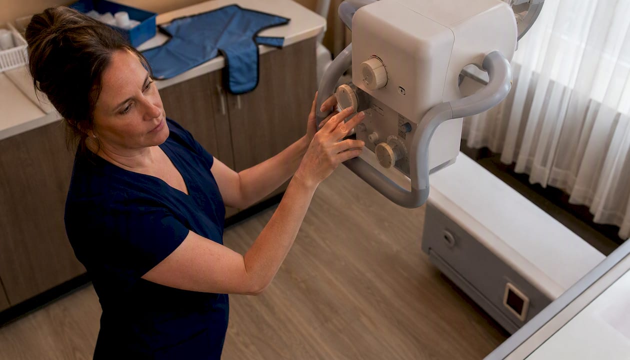
Fluoroscopy is a real-time X-ray procedure that produces continuous, movie-like images of your internal structures as they move. Unlike a standard X-ray, which captures a single still image, fluoroscopy lets your radiologist watch organs, tissues, and devices in motion. It is used to evaluate the digestive, cardiovascular, urinary, and musculoskeletal systems, and to guide procedures such as catheter placement or joint injections.
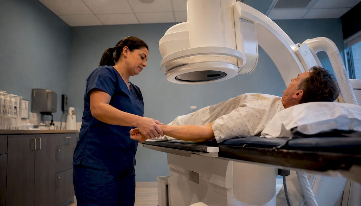
In Canada, fluoroscopy is a regulated procedure. Equipment and practice standards are set by Health Canada’s Safety Code 35, and every exam is performed under radiologist or physician supervision. Radiation dose is kept as low as reasonably achievable using modern dose-reduction techniques.
Common uses at a glance:
- Barium swallow and upper GI studies
- Angiography and vascular procedures
- Catheter and stent placement
- Guided injections for pain management
- Joint arthrograms and musculoskeletal studies
Table of Contents
- What is fluoroscopy and when does a doctor order it?
- How does fluoroscopy work?
- What are the most common fluoroscopy procedures?
- How should you prepare for a fluoroscopy exam?
- What happens during the procedure?
- What are the risks, and how is safety managed in Canada?
- Why do clinicians choose fluoroscopy over other imaging?
- How do you get your results, and what happens next?
- How does fluoroscopy compare to CT and plain X-ray?
- What Canadian patients need to know about regulations and booking
- Key takeaways
- A note from the clinical team
- Fluoroscopy at Canadian Diagnostic Network
- Useful sources and further reading
- FAQ
What is fluoroscopy and when does a doctor order it?
A standard X-ray is useful for a quick snapshot, but some clinical questions require more. When your doctor needs to see how something moves, or needs live guidance to place a device accurately, a static image is not enough.
Fluoroscopy produces real-time images by passing a continuous or pulsed X-ray beam through the body and displaying the result on a monitor. This lets the clinical team observe function, not just anatomy. A barium swallow study, for example, shows the mechanics of swallowing in motion, which a plain film simply cannot capture. A catheter placement procedure uses live imaging to confirm the device is positioned correctly before the patient leaves the table.
Clinicians typically order fluoroscopy when:
- A dynamic process needs to be evaluated (swallowing, bowel movement, joint mechanics)
- A procedure requires real-time guidance (catheter insertion, guided injection, stent deployment)
- A contrast study is needed to outline a hollow organ or vessel
The exam is ordered across multiple body systems: digestive, urinary, reproductive, vascular, and musculoskeletal. It is performed in outpatient diagnostic clinics for most routine studies, and in hospital interventional suites for complex vascular or cardiac procedures.
How does fluoroscopy work?
The core technology is a continuous or pulsed X-ray beam directed through the body toward a detector. Older systems used an image intensifier tube to amplify the signal; modern suites increasingly use digital flat-panel detectors, which produce sharper images at lower doses.
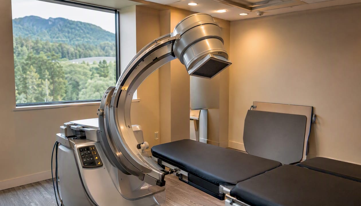
The live image appears on a monitor in the room, allowing the radiologist or proceduralist to watch in real time. When the beam is pulsed rather than continuous, the patient receives radiation only during brief intervals, which meaningfully reduces total exposure. Modern fluoroscopy suites also incorporate grid-controlled pulsed fluoroscopy, multi-beam hardening filters, and virtual collimation to further lower dose while maintaining image quality.
Contrast agents
Many fluoroscopy procedures use a contrast agent to make structures visible that would otherwise blend into surrounding tissue.
- Barium sulphate is a thick, chalky liquid swallowed or administered rectally to outline the digestive tract.
- Iodine-based contrast is injected intravenously or directly into a joint or vessel to highlight blood vessels, urinary structures, or joint spaces.
- Air is occasionally used as a negative contrast agent in certain bowel studies.
The choice of contrast depends on the organ being studied and whether the patient has any known allergies.
Pro Tip:If you have ever had a reaction to contrast dye, iodine, or shellfish, tell the booking team when you schedule your appointment, not just on the day of the exam. This gives the team time to plan any necessary precautions in advance.
What are the most common fluoroscopy procedures?
Most patients are referred for one of a handful of well-established exams. Here is what each one looks for and where it is typically performed.
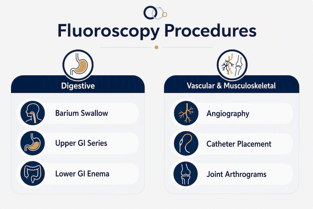
Barium swallow and upper GI series: These studies evaluate swallowing mechanics and the oesophagus, stomach, and upper small bowel. They are commonly ordered for persistent heartburn, difficulty swallowing, or suspected ulcers. Most are done in outpatient diagnostic clinics.
Lower GI (barium enema): Barium is introduced rectally to outline the colon. This study can identify polyps, narrowing, or inflammatory changes. It is less common now that colonoscopy is widely available, but still used in specific clinical situations.
Angiography and vascular studies: Iodine-based contrast is injected into blood vessels to assess flow, identify blockages, or guide interventions such as stent placement. These procedures are typically performed in hospital interventional suites with a specialist team.
Catheter and stent placement: Live imaging confirms correct device position during placement. This applies to central venous catheters, biliary stents, and ureteral stents, among others.
Guided injections for pain management: Fluoroscopy is widely used in Canada for musculoskeletal and spine interventions. Real-time needle visualisation improves placement accuracy and reduces the risk of inadvertent injury to surrounding structures.
Joint arthrograms: Contrast is injected directly into a joint space, then imaged to assess cartilage, ligaments, or the joint capsule. These are often performed in outpatient clinics and are frequently followed by MRI for detailed soft-tissue assessment.
How should you prepare for a fluoroscopy exam?
Preparation varies by exam type, but the following steps apply to most fluoroscopy appointments. Your referring clinician or the imaging clinic will give you specific instructions when you book.
- Fast if required. Upper GI and barium studies typically require fasting for four to six hours beforehand. Lower GI studies may require a bowel preparation the day before. Guided injections and joint studies generally do not require fasting.
- Review your medications. If you take blood thinners (such as warfarin or a direct oral anticoagulant), ask your doctor whether to hold the dose before an interventional procedure. Most routine diagnostic studies do not require medication changes.
- Bring your requisition and health card. Your physician’s requisition is required to book and to bill provincial health insurance where applicable. Bring photo ID and your provincial health card.
- Disclose all allergies. Tell the clinic about any allergy to iodine, shellfish, or previous contrast reactions. This is especially important if your exam involves iodine-based contrast dye.
- Inform the team if you are pregnant or may be pregnant. Radiation can harm a developing baby. Pregnancy is a contraindication for most fluoroscopy exams unless the clinical benefit clearly outweighs the risk. Bring childcare arrangements if needed.
- Arrange a ride home if sedation is planned. Most routine fluoroscopy exams do not require sedation, but interventional procedures sometimes do. Confirm this when you book.
- Wear comfortable, loose clothing. You will likely be asked to change into a gown and remove jewellery or metal objects from the area being imaged.
What happens during the procedure?
When you arrive, a technologist will review your requisition, confirm your identity, and ask about allergies and relevant medical history. You will sign a consent form before any contrast or interventional procedure begins.
You will change into a gown and remove any metal objects, including jewellery, belts, and underwire bras, from the area being examined. The technologist will position you on the imaging table, which may be tilted during the exam to move contrast through the body or to get different views.
The X-ray tube and detector are positioned around you. For most studies, you will be asked to hold still or follow specific instructions, such as swallowing on cue during a barium study. The radiologist or technologist monitors the live image on a screen in the room or from a shielded control area.
If contrast is used, it will be administered at the appropriate point in the exam, either by swallowing, enema, injection, or intravenous line, depending on the study. You may feel warmth or a brief metallic taste if iodine contrast is injected intravenously. This is normal and passes quickly.
Most outpatient fluoroscopy appointments take a limited amount of time from check-in to completion, with only a portion of that time spent under active imaging. Interventional procedures, such as vascular studies or stent placements, can take considerably longer and may involve a recovery period afterward.
What are the risks, and how is safety managed in Canada?
Radiation exposure is the primary risk associated with fluoroscopy. The dose is higher than a single plain X-ray because the beam runs continuously or in pulses over the course of the exam. That said, the exposure from a routine diagnostic fluoroscopy study is generally well within accepted safety limits.
Health Canada’s Safety Code 35 sets the framework for safe fluoroscopy practice in Canada. It requires that exams be clinically justified before they are performed, that image intensification be used, and that entrance exposure rates remain within defined limits.
| Parameter | Safety Code 35 / Equipment Standard |
|---|---|
| Maximum entrance exposure rate | Not to exceed 10 roentgens per minute at shortest target-to-skin distance |
| Typical modern equipment rate | Less than 5 roentgens per minute in most clinical settings |
| Guiding principle | ALARA (as low as reasonably achievable) |
| Dose-reduction techniques | Pulsed fluoroscopy, collimation, last-image hold, lowest acceptable frame rates |
The ALARA principle means that technologists and radiologists actively choose settings that minimise your exposure while still producing a diagnostic image. In practice, this means using pulsed rather than continuous fluoroscopy, tightly collimating the beam to the area of interest, and using the last-image hold feature to review a still frame without additional radiation.
Pro Tip:Ask your technologist whether pulsed fluoroscopy is being used for your exam. Most modern Canadian clinics use it as standard, but it is a reasonable question that shows you are engaged in your own care.
Beyond radiation, the other main risks are:
- Contrast allergy: Reactions to iodine-based contrast range from mild (hives, nausea) to rare but serious (anaphylaxis). Clinics have protocols and medications on hand to manage reactions.
- Kidney considerations: Iodine contrast can affect kidney function in patients with pre-existing renal disease. Your team will review your history and may check kidney function before proceeding.
- Pregnancy: Radiation exposure during pregnancy carries risk to the developing baby. Fluoroscopy is avoided in pregnancy unless the benefit clearly outweighs the risk, and the team will ask about pregnancy status before every exam.
- Higher-dose modes: Cinefluorography (cine mode) uses higher tube currents and voltages and increases patient dose compared with standard fluoroscopy. It is used only when its diagnostic benefit justifies the additional exposure.
For context on cumulative radiation exposure across multiple imaging studies, your radiologist or referring physician can advise based on your individual history.
Why do clinicians choose fluoroscopy over other imaging?
The defining advantage of fluoroscopy is real-time visualisation. No other widely available modality lets a clinician watch a needle advance into a joint, confirm a catheter tip position, or observe the exact moment a patient’s swallowing mechanism fails.
Key clinical benefits include:
- Dynamic assessment: Conditions that only manifest during movement, such as swallowing dysfunction or intermittent bowel obstruction, are often invisible on static imaging.
- Procedural guidance: Live imaging reduces the risk of misplacement during interventional procedures, which can mean fewer complications and less need for repeat procedures.
- Immediate decision-making: The radiologist can adjust the procedure in real time based on what the image shows, rather than waiting for a report.
- Diagnosis and treatment in one session: Many interventional fluoroscopy procedures both identify and address the problem during the same appointment, such as placing a stent to relieve a biliary obstruction.
The justification principle in Safety Code 35 reinforces this: fluoroscopy should only be used when it provides unique, real-time information that a standard X-ray or other modality cannot capture.
How do you get your results, and what happens next?
After your exam, the images are reviewed by a radiologist, a physician who specialises in interpreting medical imaging. The radiologist prepares a written report that includes findings, an overall impression, and any recommendations for follow-up.
- Turnaround time: For routine outpatient exams, many Canadian diagnostic centres provide reports within 24–48 hours. Urgent or time-sensitive findings are communicated to the referring clinician immediately.
- Who receives the report: Your referring physician receives the report directly. You can request a copy through the CDN patients portal or by contacting the clinic.
- What the report contains: The radiologist describes what was seen, notes any abnormalities, and may suggest additional imaging or clinical follow-up if warranted.
- Next steps: Your referring clinician will discuss the results with you and recommend any further investigations or treatment. If your symptoms worsen before your follow-up appointment, contact your doctor or the clinic directly.
How does fluoroscopy compare to CT and plain X-ray?
Understanding the difference between fluoroscopy and a standard X-ray helps you understand why your doctor chose this particular test for your situation.
| Modality | What it shows | Radiation dose | Best used for |
|---|---|---|---|
| Plain X-ray | Static 2D image of dense structures | Low (single exposure) | Fractures, chest screening, quick structural checks |
| Fluoroscopy | Real-time moving images of organs and devices | Moderate (continuous or pulsed beam) | Dynamic studies, procedural guidance, contrast studies |
| CT scan | Cross-sectional 3D detail of all tissue types | Higher (multiple X-ray passes) | Complex anatomy, tumour staging, trauma assessment |
A plain X-ray is the right choice when a quick structural snapshot is all that is needed. CT provides the most anatomical detail and is preferred for complex diagnostic questions involving soft tissue, vessels, or three-dimensional relationships. Fluoroscopy fills the gap when movement or real-time guidance is the clinical priority.
In some cases, the two modalities are used together. A CT scan may be used for pre-procedure planning, with fluoroscopy providing live guidance during the intervention itself.
What Canadian patients need to know about regulations and booking
Fluoroscopy in Canada is governed by two overlapping frameworks. The Radiation Emitting Devices Act (REDA) is federal legislation that sets equipment safety standards for all radiation-emitting devices, including fluoroscopy units. Health Canada’s Safety Code 35 builds on REDA by providing detailed operational guidance for medical facilities: equipment specifications, dose limits, justification requirements, and quality assurance protocols.
While REDA and federal standards set the baseline, provinces maintain their own licensing requirements for radiological technologists and clinical protocols for use. This means your experience may vary slightly depending on where in Canada you receive care, but the core safety standards apply nationally.
What to expect when booking in Ontario:
- You need a physician’s requisition. Fluoroscopy is not available as a walk-in service; your family doctor or specialist must refer you.
- In Ontario, many fluoroscopy procedures are covered by OHIP when medically necessary and performed at an accredited facility. Some specialised or interventional procedures may involve additional fees; confirm coverage with your referring clinician and the clinic when you book.
- Technologists performing fluoroscopy in Ontario are registered with the College of Medical Radiation and Imaging Technologists of Ontario (CMRITO) and work under radiologist supervision.
- Canadian Diagnostic Network (CDN) operates more than 20 clinic locations in the Ottawa area, with radiologist oversight and modern dose-reduction equipment at each site. You can book by contacting the clinic directly after receiving your requisition.
Key takeaways
Fluoroscopy is a real-time X-ray procedure that is safe, regulated under Health Canada’s Safety Code 35, and used across a wide range of diagnostic and interventional applications in Canada.
| Point | Details |
|---|---|
| What fluoroscopy is | A real-time X-ray that produces moving images to evaluate organ function and guide procedures. |
| Regulated in Canada | Health Canada’s Safety Code 35 and REDA set dose limits, equipment standards, and justification requirements. |
| Dose is actively minimised | ALARA, pulsed fluoroscopy, and collimation keep exposure well below the 10 roentgens per minute maximum. |
| What to tell your team | Disclose all allergies (especially iodine or contrast), pregnancy status, and current medications before your exam. |
| Cdncare in Ottawa | Canadian Diagnostic Network offers fluoroscopy across more than 20 Ottawa-area clinics with radiologist oversight and 24–48 hour report turnaround. |
A note from the clinical team
Many patients arrive for fluoroscopy having already had a plain X-ray or ultrasound that did not give their doctor a complete picture. A common scenario: someone referred after months of swallowing difficulty, where a barium swallow finally captures the exact moment the problem occurs. Another: a patient with chronic back pain who has tried conservative management and now needs a fluoroscopically guided injection to confirm the pain source and deliver targeted treatment.
What patients often tell us is that they were not expecting the procedure to be as straightforward as it was. Most routine fluoroscopy exams involve no sedation, no significant discomfort, and a short time in the room. The contrast drink or injection is the part that surprises people most, and even that is usually well tolerated.
We ask every patient to bring their requisition, health card, and a list of current medications. If you have had a contrast reaction before, let us know when you call to book. We will review your history, flag it for the radiologist, and make sure the right precautions are in place before you arrive.
*— Felix
Fluoroscopy at Canadian Diagnostic Network
Getting a fluoroscopy referral can feel like a lot to navigate, particularly if it is your first time. At Canadian Diagnostic Network, the process is designed to be straightforward. Once your physician has issued a requisition, you contact one of our Ottawa-area clinic locations to book your appointment. Our team will walk you through any preparation steps specific to your exam and answer questions about what to expect on the day.
CDN’s fluoroscopy suites use modern dose-reduction equipment and operate under direct radiologist supervision. Reports for routine outpatient exams are typically ready within 24–48 hours and go directly to your referring clinician. For patients who need faster access, our urgent care imaging service offers walk-in X-ray and same-day options across the Ottawa region.
To book, contact your nearest CDN location after receiving your physician’s requisition.
Useful sources and further reading
The following sources provide authoritative guidance on fluoroscopy, radiation safety, and patient preparation. Reviewing them alongside a conversation with your referring clinician will give you the most complete picture for your specific situation.
- Health Canada Safety Code 35: The national standard for X-ray equipment installation, use, and dose limits in Canadian medical facilities, including fluoroscopy.
- Canada.ca: Medical and dental X-rays: Plain-language overview of how X-ray and fluoroscopy imaging works and what body systems it covers.
- CDC: Facts about fluoroscopy: A concise summary of fluoroscopy uses, risks, and dose considerations from the U.S. Centers for Disease Control and Prevention.
- Government of Canada: X-ray equipment safety procedures: The full technical publication behind Safety Code 35, including entrance exposure rate limits and equipment requirements.
- Canada.ca: Government’s role in radiation protection: Explains the division of responsibility between federal (REDA, Safety Code 35) and provincial (licensing, clinical protocols) oversight.
- MedlinePlus: Fluoroscopy: A patient-facing reference from the U.S. National Library of Medicine covering what fluoroscopy is, how it works, and what to expect.
- CDN patients portal: Access your results, manage appointments, and find preparation documents for your CDN imaging exam.
Discuss your specific results and any follow-up steps with your referring clinician. This article provides general information and is not a substitute for personalised medical advice.
FAQ
What is fluoroscopy and why is it used?
Fluoroscopy is a real-time X-ray that produces moving images of internal structures, allowing clinicians to evaluate organ function or guide procedures such as catheter placement and contrast studies. It is ordered when a static image is not sufficient to answer the clinical question.
Are you awake during a fluoroscopy?
Yes, most fluoroscopy exams are performed while you are fully awake. Sedation is occasionally used for longer interventional procedures, but routine diagnostic studies such as barium swallows or guided injections do not require it.
How long does a fluoroscopy procedure take?
Most outpatient fluoroscopy appointments take a limited amount of time from check-in to completion, with only a portion of that time spent under active imaging. Interventional procedures can take longer and may include a recovery period.
What is the difference between a CT scan and fluoroscopy?
CT produces detailed cross-sectional, three-dimensional images of all tissue types and is best for complex anatomy or tumour assessment. Fluoroscopy provides real-time moving images and is preferred when dynamic function or live procedural guidance is the clinical priority.
Recommended
Your examination or doctor’s visit is 4 easy steps away