We now offer x-ray appointments at our Phenix, Charlotte, Rideau Valley, Nepean, Kanata, and Hunt Club clinics. Please check location availability before visiting.

Request an Appointment
Why imaging matters in oncology: a practical guide

TL;DR:
- Imaging is vital throughout cancer care, guiding diagnosis, staging, treatment, and monitoring. It often alters management decisions, replacing invasive procedures and refining treatment pathways. Canadian programs like Cdncare offer accessible, rapid, and guideline-supported imaging services across Ottawa.
Diagnostic imaging is essential at every stage of cancer care. It locates disease, defines how far it has spread, guides treatment decisions, and measures whether therapy is working. Without it, clinicians would be making critical decisions without the evidence they need. A PET/CT scan that reveals unsuspected lymph node involvement in a patient thought to have early-stage lymphoma can shift the entire treatment plan from local radiotherapy to systemic chemotherapy. A screening mammogram through the Ontario Breast Screening Program can catch a tumour before it causes symptoms, when treatment is most likely to succeed.
The short version: imaging changes management. It upgrades or downgrades stage, redirects therapy, and often replaces the need for open surgical biopsy.
Three clinical impacts worth knowing from the start:
- Staging accuracy: Imaging, particularly PET/CT, routinely reveals disease extent that physical examination and blood tests miss, changing the treatment pathway.
- Treatment guidance: Radiotherapy planning, surgical resection margins, and interventional procedures all depend on precise anatomical and functional imaging.
- Response assessment: Structured criteria like RECIST and PERCIST give clinicians an objective way to decide whether to continue, switch, or intensify therapy.
Table of Contents
- How imaging guides you through every stage of cancer care
- What does each imaging modality actually show you?
- How does imaging measure whether treatment is working?
- Why imaging is central to oncology clinical trials
- What are the real limitations and risks of oncology imaging?
- Navigating cancer imaging in Canada: guidelines, programmes, and access
- Key takeaways
- What clinicians and patients often underestimate about imaging
- Cdncare's cancer imaging services in Ottawa
- Useful sources and guidelines
- FAQ
How imaging guides you through every stage of cancer care
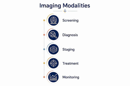
The cancer care continuum runs from initial suspicion through screening, diagnosis, staging, treatment, monitoring, and long-term survivorship. Imaging has a defined role at each point, and understanding where you are in that continuum helps you know what to expect from each scan.
Screening: catching cancer before symptoms appear
Screening is population-level imaging in people without symptoms. The clearest Canadian example is mammography. The Ontario Breast Screening Program offers screening mammography to women aged 50-74 at average risk, and to women aged 30 and older at high risk. Screening mammography is specifically designed to detect small, non-palpable tumours. It is not the same as a diagnostic mammogram ordered because of a lump or nipple discharge. Understanding that distinction matters: a screening result that prompts a callback is not a cancer diagnosis; it is a signal that a closer look is warranted.
Pro Tip: If you are a clinician referring patients for breast screening, confirm eligibility under the OBSP before ordering a diagnostic mammogram - the pathway, reporting standard, and follow-up protocol differ significantly between the two.
Real-world screening catches cancers that patients would not have noticed for months or years. The importance of regular mammography is well documented: earlier detection consistently correlates with a broader range of treatment options and better outcomes.
Diagnosis and localisation: from suspicion to confirmation
Once a lesion is identified, imaging shifts from population screening to individual diagnosis. CT, MRI, and ultrasound characterise the lesion: its size, borders, internal structure, and relationship to surrounding tissue. This information tells the clinical team whether a biopsy is needed, and if so, where to target it.
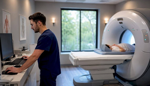
Interventional radiology has changed biopsy practice significantly. Image-guided biopsy, using ultrasound or CT for real-time needle guidance, reduces the need for open surgical biopsy in many cases, shortens time to tissue diagnosis, and lowers complication rates. For a patient with a deep retroperitoneal mass, a CT-guided biopsy under local anaesthetic is far less disruptive than an open procedure.
Staging: defining how far disease has spread
Staging is where imaging most directly determines the treatment pathway. A patient staged as having localised disease may be a surgical candidate; the same patient with distant metastases may not be. PET/CT is particularly valuable here because it combines metabolic activity (from the PET component) with anatomical detail (from the CT), detecting sites of disease that CT alone can miss.
Canadian clinical practice guidelines recommend PET/CT for staging and restaging in certain cancers, including lymphoma, because it provides functional information that can change treatment decisions when anatomical imaging is inconclusive. For instance, in diffuse large B-cell lymphoma, PET/CT has high sensitivity for bone marrow involvement, often replacing the need for bone marrow biopsy.
Treatment planning and monitoring
Radiotherapy planning relies on CT and MRI to define the target volume and the surrounding organs at risk. Adaptive radiotherapy uses repeat imaging during a treatment course to adjust for tumour shrinkage or patient anatomy changes. Surgical planning uses MRI to map tumour proximity to critical structures.
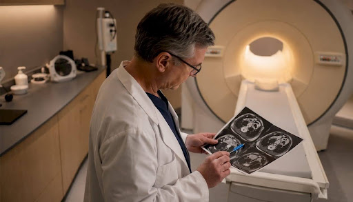
After treatment begins, response imaging tells the team whether the plan is working. Routine surveillance imaging after treatment completion looks for recurrence, though the frequency and modality depend on cancer type and applicable guidelines. Many patients ask for more frequent scans than guidelines recommend, often out of anxiety rather than clinical need. Addressing that expectation directly, and explaining what each scan is designed to detect, reduces unnecessary radiation exposure and helps patients feel more in control.
What does each imaging modality actually show you?
Choosing the right modality means matching the clinical question to what each technology can answer. Here is a practical breakdown.
Mammography
Mammography uses low-dose X-ray to image breast tissue. It detects microcalcifications and soft-tissue densities that are often the earliest signs of breast cancer. Its main limitation is reduced sensitivity in dense breast tissue, where ultrasound or MRI may add value. Digital mammography and tomosynthesis (3D mammography) improve detection in dense breasts compared to standard 2D imaging.
Ultrasound
Ultrasound for cancer uses sound waves and produces no ionising radiation, making it safe for repeated use and for younger patients. It is particularly useful for characterising breast lesions found on mammography, guiding biopsies, and evaluating superficial lymph nodes. Its limitation is operator dependence and reduced ability to image through bone or air.
CT (computed tomography)
CT provides rapid, detailed cross-sectional images of the chest, abdomen, and pelvis. It is the workhorse of oncology staging and surveillance. CT is fast, widely available, and excellent for detecting pulmonary metastases and lymphadenopathy. The main limitation is ionising radiation and the use of iodinated contrast, which carries a small risk of allergic reaction and nephrotoxicity in patients with impaired kidney function.
MRI (magnetic resonance imaging)
MRI offers superior soft-tissue contrast without ionising radiation. It is the preferred modality for brain tumours, spinal cord involvement, pelvic malignancies (rectal, cervical, prostate), and liver characterisation. Functional MRI sequences, including diffusion-weighted imaging, add information about tumour cellularity and treatment response. Limitations include longer scan times, claustrophobia, contraindications for patients with certain metallic implants, and limited availability in some regions.
Whole-body MRI (WB-MRI) can stage multiple myeloma and other cancers in a single session without ionising radiation. Patient preference studies show many patients favour WB-MRI over conventional staging pathways if accuracy and time to diagnosis are comparable, though long scan times and claustrophobia remain practical barriers.
PET/CT
PET/CT combines a metabolic tracer (most commonly 18F-FDG, which accumulates in metabolically active tissue) with CT anatomy. It detects sites of disease that are metabolically active but not yet large enough to appear abnormal on CT alone. Evidence from systematic reviews confirms that 18F-FDG PET/CT has high sensitivity and specificity for detecting bone marrow involvement in diffuse large B-cell lymphoma and improves staging accuracy in non-small cell lung cancer. PSMA PET/CT shows high specificity for prostate cancer bone metastasis.
Pro Tip: PET/CT adds the most value when the clinical question cannot be answered by CT or MRI alone - for example, when CT shows a residual mass after lymphoma treatment and you need to know whether it is viable tumour or fibrosis. Adding PET/CT to the pathway in that scenario is guideline-supported and avoids unnecessary salvage therapy.
SPECT and nuclear medicine
SPECT (single-photon emission computed tomography) uses gamma-emitting radiotracers to image organ function and detect bone metastases. Bone scintigraphy remains widely used for detecting skeletal spread in breast and prostate cancer. SPECT/CT combines functional and anatomical data, improving specificity over planar scintigraphy alone.
Interventional radiology
Interventional radiology sits at the intersection of diagnosis and treatment. Image-guided procedures include biopsy, tumour ablation (radiofrequency, microwave, cryoablation), hepatic arterial embolisation, and nephrostomy. These procedures are less invasive than surgery, often performed under local anaesthetic, and can be both diagnostic and therapeutic in the same session.
According to the Canadian Medical Imaging Inventory, Canada performed a large volume of CT exams in the 2022-2023 reporting period, with a significant portion attributed to oncology indications. PET/CT volumes were also substantial, with the majority used for oncology purposes.
How does imaging measure whether treatment is working?
Response assessment is one of the most clinically consequential uses of imaging, and it depends on applying the right criteria at the right time.
Response criteria: RECIST, PERCIST, and beyond
| Criteria | What it measures | Key clinical use |
|---|---|---|
| RECIST 1.1 | Change in tumour size (longest diameter of target lesions) | Standard for solid tumours in clinical trials and routine practice |
| PERCIST | Change in FDG uptake on PET/CT (standardised uptake value) | Lymphoma, lung cancer, and other FDG-avid tumours |
| Lugano criteria | Combined size and metabolic response (Deauville score) | Lymphoma response assessment |
| PI-RADS | Prostate lesion risk stratification on MRI | Prostate cancer detection and biopsy targeting |
| BI-RADS | Breast lesion risk stratification on mammography/ultrasound | Breast cancer screening and diagnostic follow-up |
RECIST 1.1 measures tumour size on CT and defines complete response, partial response, stable disease, and progressive disease by percentage change in target lesion diameter. PERCIST measures metabolic activity and is more sensitive for detecting early response in FDG-avid tumours, sometimes showing response weeks before CT demonstrates size change. The Lugano criteria, which incorporate the five-point Deauville scale for PET response, are the standard for lymphoma.
Timing matters as much as the criteria
Scanning too early after chemotherapy can produce false-positive results due to treatment-related inflammation. Scanning too late delays a management decision. A general framework:
- Early response assessment: typically after 2-4 cycles of chemotherapy, using PET/CT for FDG-avid tumours or CT for solid tumours
- End-of-treatment assessment: 6-8 weeks after completing chemotherapy or radiotherapy, to allow treatment-related changes to settle
- Surveillance imaging: interval and modality determined by tumour type and applicable guidelines; not every cancer warrants routine surveillance CT
Pitfalls: pseudoprogression and treatment-related inflammation
Pseudoprogression, where tumours appear to enlarge or show increased FDG uptake early in immunotherapy, can mimic true progression on both anatomical and functional imaging. Treatment-related inflammation after radioembolisation or immunotherapy produces similar appearances. The practical guidance: when imaging and clinical trajectory are discordant, correlate carefully and consider follow-up imaging or biopsy before changing therapy. Switching treatment based on a single ambiguous scan is a common and avoidable error.
Why imaging is central to oncology clinical trials
Clinical trials in oncology depend on imaging at every step: eligibility confirmation, stratification, and the primary endpoints that determine whether a drug works.
How imaging defines trial eligibility and endpoints
Most solid tumour trials require measurable disease by RECIST 1.1 at enrolment. Patients without measurable lesions on baseline imaging may be ineligible, regardless of clinical evidence of disease. Imaging endpoints, including objective response rate and progression-free survival, are the primary measures of efficacy in most phase II and many phase III trials.
Common operational challenges in trial imaging include:
- Protocol variability: different CT slice thicknesses, contrast protocols, and scan timing across sites introduce measurement variability that can obscure real treatment effects.
- Workflow delays: late or missed scans create missing data that complicate per-protocol analyses.
- Inter-reader variability: different radiologists applying RECIST to the same scan can reach different conclusions, particularly for borderline lesions.
- Equipment heterogeneity: PET/CT scanner differences in sensitivity and reconstruction algorithms affect SUV measurements used in PERCIST.
Practical recommendations for trial imaging reliability
Standardised imaging protocols, specified in the trial protocol and enforced by a central imaging core laboratory, reduce inter-reader variability and improve endpoint reliability. Centralised imaging review is often the difference between a usable and an unusable imaging endpoint. Pre-specified imaging timelines, with windows that account for treatment delays, prevent the missing-data problems that compromise per-protocol analyses.
The scale of oncology imaging in Canada underscores why standardisation matters: with more than 65% of PET/CT exams performed for oncology indications nationally, even small protocol variations across sites accumulate into meaningful measurement error at the trial level.
What are the real limitations and risks of oncology imaging?
Imaging is powerful, but it is not infallible. Being honest about its limits helps you counsel patients and avoid low-value testing.
Key risks to discuss with patients
- Ionising radiation: CT and PET/CT involve radiation exposure. A single CT of the chest, abdomen, and pelvis delivers a meaningful dose. Cumulative exposure across multiple scans over a cancer treatment course warrants consideration, particularly in younger patients.
- Contrast reactions: Iodinated CT contrast and gadolinium MRI contrast both carry small risks of allergic reaction. Gadolinium deposition in brain tissue with repeated MRI contrast use is an area of ongoing study.
- False positives and overdiagnosis: Screening programmes detect some cancers that would never have caused symptoms or death. Incidental findings on staging CT, such as adrenal nodules or pulmonary nodules, trigger additional imaging and sometimes biopsy, with associated anxiety and cost.
- False negatives: No modality is 100% sensitive. A normal scan does not rule out disease, particularly micrometastatic disease below the resolution threshold of current technology.
- Access delays: Wait times for CT and MRI in Canada have increased between 2012 and 2023 per CMII data. Delays in diagnostic imaging translate directly into delays in treatment initiation.
Pro Tip: When a patient asks "Can't I just have a scan to be sure?", the most helpful response is to explain what the scan is designed to detect and what it cannot rule out. A negative CT does not mean no cancer; it means no cancer visible at CT resolution on that day.
When to prioritise urgent imaging
Rapid clinical change, suspected spinal cord compression, new neurological signs in a patient with known malignancy, and discordant test results (for example, rising tumour markers with a stable-appearing CT) are all indications for urgent or repeat imaging. In these situations, waiting for a routine appointment is not appropriate.
Patients often report that wait times and difficulty interpreting reports are among their greatest sources of anxiety during cancer care. Clear communication about what a scan can and cannot tell you, and realistic timelines for results, goes a long way toward reducing that anxiety.
Navigating cancer imaging in Canada: guidelines, programmes, and access
Canada has national and provincial resources that shape how oncology imaging is accessed and delivered.
Canadian guidelines and PET/CT recommendations
Canadian clinical practice guidelines support PET/CT for staging and restaging in lymphoma, and evidence supports its use in non-small cell lung cancer staging and PSMA PET/CT for prostate cancer. The All.Can Canada report recommends that imaging be part of a standardised care pathway, with rapid, accurate imaging and clear communication to reduce delays to treatment. Economic evidence suggests PET/CT can be cost-effective for staging early-stage follicular lymphoma and non-small cell lung cancer, though surveillance PET/CT is not consistently cost-effective compared to CT surveillance.
National imaging volumes and oncology proportions
| Modality | National exam volume (2022-2023) | Oncology proportion |
|---|---|---|
| CT | 6,385,665 | ~25% |
| PET/CT | 156,320 | >65% |
| SPECT | 929,010 | Not specified |
Source: Canadian Medical Imaging Inventory 2022-2023
Practical access: OBSP, provincial programmes, and CDN
The Ontario Breast Screening Program provides organised, funded breast screening for eligible Ontario residents. Cdncare operates one of the largest OBSP networks in Ontario, with mammography services and breast ultrasound available across more than 20 clinic locations in Ottawa.
Access considerations for patients and referring clinicians:
- Wait times: CT and MRI wait times have increased nationally. For non-urgent oncology imaging, plan for potential delays and build that into your referral timeline.
- Rural and remote access: PET/CT is concentrated in major urban centres. Patients in rural areas may require travel or referral to a regional cancer centre for PET/CT access.
- Referral pathways: Provincial cancer programmes and regional cancer centres coordinate PET/CT access for eligible patients. Confirm provincial criteria before referring, as funded indications vary by province.
- Turnaround times: Cdncare delivers results within 24-48 hours for most studies, supporting faster clinical decisions.
Key takeaways
Imaging is the single most important non-invasive tool in cancer care, shaping decisions from first suspicion through long-term survivorship.
| Point | Details |
|---|---|
| Imaging changes management | PET/CT, CT, and MRI routinely alter staging and redirect therapy in ways that physical examination cannot. |
| Match modality to the question | Each scan answers a specific clinical question; choosing the wrong modality wastes time and may miss the answer you need. |
| Follow guideline-driven timing | Scanning too early or too late produces misleading results; response imaging has defined windows for a reason. |
| Communicate clearly with patients | Patients value timely results and plain-language explanations; address wait-time anxiety and report interpretation directly. |
| Cdncare provides accessible oncology imaging | With over 20 Ottawa locations, OBSP participation, and 24-48 hour turnaround, Cdncare supports faster diagnosis and treatment planning. |
What clinicians and patients often underestimate about imaging
There is a tendency, understandable on both sides of the consultation, to treat imaging as a binary: either the scan shows something or it does not. The reality is more nuanced, and that nuance has real consequences.
The most common gap we see is not in the technology but in how results are communicated. Imaging reports are written for radiologists and oncologists, not for patients sitting at home trying to make sense of a PDF. A report that says "no evidence of disease progression" is reassuring to a clinician who knows what progression means in that context. To a patient who has been anxious for two weeks, the same sentence can feel ambiguous or even alarming. Translating that finding into a clear management statement, "your scan shows no new disease; we will continue your current treatment and repeat imaging in three months," takes thirty seconds and changes the patient's experience of their care.
The second underestimated issue is the expectation gap around surveillance imaging. Many patients want more frequent scans than guidelines recommend, often because they equate scanning with safety. Addressing this directly, explaining what the scan is designed to detect, what it cannot rule out, and why more frequent scanning carries its own risks, is one of the most valuable conversations a clinician can have. It is also one of the most frequently skipped.
Cdncare's cancer imaging services in Ottawa
Cdncare is Ottawa's largest diagnostic imaging provider, with more than 20 clinic locations and a team of over 150 radiologists and technologists. For patients and clinicians navigating cancer care, that breadth of access matters: you are not waiting weeks for a single appointment at a single site.
Relevant services for oncology patients and referring clinicians include:
- Mammography and OBSP: Cdncare participates in the Ontario Breast Screening Program. You can book a screening or diagnostic mammogram directly, with results typically available within 24-48 hours.
- Breast ultrasound: Available for characterising mammography findings, evaluating dense breast tissue, and guiding biopsy. Book through the breast ultrasound service page.
- Diagnostic ultrasound: Useful across multiple oncology indications, from lymph node assessment to abdominal organ evaluation.
- Image-guided procedures: CDN collaborates with hospital and specialist partners for image-guided biopsy and interventional procedures where required.
For urgent diagnostic needs, walk-in ultrasound and X-ray is available without a long wait. To book a mammogram or confirm OBSP eligibility, visit the Cdncare mammography page or contact your nearest clinic directly.
Useful sources and guidelines
The following sources are worth bookmarking if you want to go deeper on any of the topics covered here.
- Imaging and cancer: A review (PMC/NIH): A peer-reviewed overview of imaging's role across the cancer care continuum; useful for clinicians wanting an evidence base for the modality discussions above.
- PET/CT capacity for cancer diagnosis and follow-up: rapid review: The most relevant Canadian evidence synthesis for PET/CT indications, cost-effectiveness, and capacity; covers lymphoma, lung, and prostate cancer in detail.
- Canadian Medical Imaging Inventory 2022-2023: National data on imaging volumes, modality distribution, and wait times; the primary source for Canadian capacity figures.
- All.Can Canada: Optimizing diagnosis in Canadian cancer care: Patient-centred recommendations on standardised diagnostic pathways and communication; directly relevant to the access and communication sections.
- Cancerpedia: Medical imaging chapter: A clear, accessible overview of imaging modalities and their roles in oncology; useful as a patient resource.
- CCSN patient survey infographic: Documents patient priorities around diagnosis, including the importance of timely results and clear communication.
- Ontario Breast Screening Program (OBSP) via Cdncare: Eligibility criteria, how to book, and what to expect from OBSP screening in Ontario.
For clinicians: consult your provincial cancer programme's referral guidelines for PET/CT funded indications, as criteria and access pathways vary by province. For patients: your family physician or oncologist can confirm which imaging is covered under your provincial plan and refer you to the appropriate programme.
"Rapid, accurate imaging and clear communication between diagnostic services and tumour teams are among the most modifiable factors in reducing time to treatment in Canadian cancer care." - All.Can Canada
FAQ
What is the role of imaging in oncology?
Imaging locates tumours, defines how far cancer has spread, guides treatment planning, measures response to therapy, and monitors for recurrence. It is used at every stage of cancer care, from screening through survivorship.
Why is PET/CT particularly valuable in cancer staging?
PET/CT combines metabolic and anatomical information, detecting disease that is active but not yet large enough to appear abnormal on CT alone. Canadian guidelines support its use in lymphoma, lung cancer, and prostate cancer staging because it frequently changes the treatment pathway.
What do BI-RADS and RECIST mean on an imaging report?
BI-RADS is a standardised scoring system for breast imaging findings, ranging from 1 (normal) to 6 (known malignancy), that tells your care team how likely a finding is to be cancerous. RECIST 1.1 is a set of criteria used to measure tumour size changes on CT during treatment, defining whether disease is responding, stable, or progressing.
How does imaging affect cancer treatment decisions?
Imaging directly determines which treatment pathway is appropriate. A staging scan that reveals distant metastases may shift a patient from a curative surgical plan to systemic therapy. A response scan showing complete metabolic remission on PET/CT may allow de-escalation of treatment.
Where can patients in Ottawa access cancer imaging?
Cdncare operates more than 20 diagnostic imaging clinics in Ottawa, including OBSP-accredited mammography, breast ultrasound, and diagnostic ultrasound services, with results typically available within 24-48 hours. Referrals for PET/CT are coordinated through provincial cancer programmes and regional cancer centres.
Recommended
- Why is Compression Important for a Mammogram? | Canadian Diagnostic Network
- Charlotte Imaging | Canadian Diagnostic Network
- Ultrasound for Cancer | Canadian Diagnostic Network
- Urgent Care Imaging | Ultrasound & X-Ray (Walk-In) | CDN
Your examination or doctor’s visit is 4 easy steps away