We now offer x-ray appointments at our Phenix, Charlotte, Rideau Valley, Nepean, Kanata, and Hunt Club clinics. Please check location availability before visiting.

Request an Appointment
What is 3D mammography and why it matters
TL;DR:
- 3D mammography improves breast cancer detection by producing layered images that better reveal abnormalities in dense tissue. It reduces recall rates and detects more invasive cancers compared to traditional 2D screening. Women with dense breasts or higher risk factors should consider its use for more effective screening.
3D mammography, known clinically as digital breast tomosynthesis, is a breast imaging technique that produces layered, three-dimensional images of breast tissue to improve cancer detection. Unlike a traditional 2D mammogram, which captures a single flat image, tomosynthesis takes multiple X-ray images from different angles and assembles them into thin, viewable slices. Studies show it detects invasive cancers 15%-22% more often than standard 2D imaging. That improvement is meaningful, particularly for women with dense breast tissue, where 2D images frequently miss tumours hiding behind overlapping tissue.
What is 3D mammography and how does the exam work?
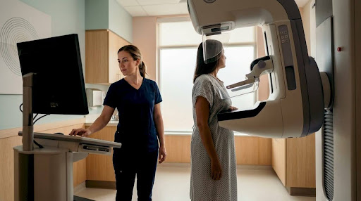
The tomosynthesis machine moves in an arc over your breast, capturing multiple X-ray images from several angles in a single pass. A computer then combines those images into a series of thin slices, similar to flipping through pages of a book. Radiologists can scroll through each layer individually, rather than reading one flat image where tissue can overlap and obscure findings.
During the exam, your breast is compressed in the same way as a standard mammogram. You hold your breath for a few seconds while the machine completes its arc. This brief breath-hold reduces motion blur and produces cleaner images. The full procedure typically takes only a few minutes longer than a conventional mammogram.
Many facilities capture both 3D and 2D images in the same appointment. This gives your radiologist a complete picture without requiring a second visit. The combined session does increase radiation exposure slightly, but doses remain within safe limits established by health regulators.
Pro Tip: Wear a two-piece outfit to your appointment. You will only need to undress from the waist up, which makes the process quicker and more comfortable.
How does 3D mammography compare to traditional 2D screening?
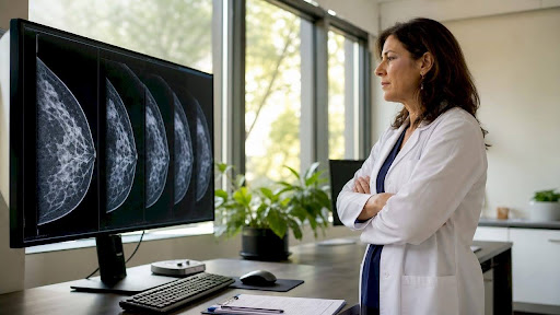
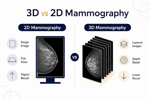
The core difference between 3D and 2D mammography comes down to image depth. A 2D mammogram compresses all breast tissue into a single flat image. When tissue layers overlap, both healthy dense tissue and potential tumours appear white, making it difficult for radiologists to distinguish one from the other.
Dense tissue masks cancers visually in 2D because both appear the same colour on the image. Tomosynthesis separates those layers, allowing radiologists to examine each slice independently and identify abnormalities with greater confidence.
"3D mammography transitions from flat 2D images to sliced, layered views, improving radiologists' ability to identify tumours clearly." - American Cancer Society
The recall rate for 2D mammography sits at 5%-12% for suspicious findings. Multiple studies show 3D mammography reduces that recall rate. Fewer callbacks mean less anxiety for patients and fewer unnecessary follow-up procedures.
| Feature | 2D mammography | 3D mammography |
|---|---|---|
| Image type | Single flat image | Multiple layered slices |
| Dense tissue performance | Limited; tissue overlaps | Improved; layers separated |
| Invasive cancer detection | Baseline | 15%-22% higher |
| Recall rate | 5%-12% | Reduced across studies |
| Radiation dose | Standard | Slightly higher, within safe limits |
Who should consider 3D mammography?
Women with dense breast tissue benefit most from 3D mammography. Approximately 40% of women have dense breast tissue, which significantly limits what a 2D mammogram can reveal. If your previous mammogram report noted dense tissue, discussing tomosynthesis with your doctor is a reasonable next step.
Beyond breast density, the following groups are strong candidates for 3D screening:
- Women aged 40 and older following routine screening guidelines
- Women with a personal or family history of breast cancer
- Women who have had inconclusive or recalled 2D mammograms in the past
- Women at elevated risk due to genetic factors such as BRCA1 or BRCA2 variants
3D mammography is used for both routine screening and diagnostic purposes. Screening applies to women with no current symptoms. Diagnostic imaging is ordered when a specific concern, such as a lump or nipple discharge, needs further evaluation. Your doctor or referring physician will specify which type of exam you need.
Insurance coverage for 3D mammography varies depending on your province, plan, and provider. Some patients face additional out-of-pocket fees compared to a standard mammogram. Confirming coverage with your insurer before your appointment avoids surprises.
What to expect before, during, and after your appointment
Knowing what happens at each stage of your appointment reduces uncertainty and helps you feel prepared.
- Scheduling. Book your mammogram when your breasts are least tender, typically in the week after your period ends. Avoid scheduling during the week before your period if possible.
- Preparation. Do not apply deodorant, antiperspirant, lotion, or powder to your underarms or chest on the day of your exam. These products can appear as white spots on the image and interfere with the reading. Review what to expect at your first mammogram if this is your first time.
- During the exam. A technologist will position your breast on the imaging platform and apply compression. The machine moves in its arc while you hold your breath briefly. Each view takes only seconds.
- After the exam. A radiologist reviews your images and sends a report to your referring physician, typically within 24-48 hours at facilities like Cdncare. If the radiologist identifies something that needs a closer look, you may be called back for additional imaging such as a breast ultrasound.
- Follow-up. A callback does not mean cancer has been found. It means the radiologist wants more information. Most callbacks result in a normal finding after additional imaging.
Pro Tip: Bring your previous mammogram images or records if you are visiting a new facility. Comparison with prior exams helps radiologists identify changes over time.
Key takeaways
3D mammography, or digital breast tomosynthesis, is the most effective standard screening tool for detecting invasive breast cancer early, particularly in women with dense breast tissue.
| Point | Details |
|---|---|
| Detection improvement | 3D mammography finds invasive cancers 15%-22% more often than 2D imaging. |
| Dense breast advantage | About 40% of women have dense tissue; 3D separates layers that 2D cannot. |
| Reduced callbacks | 3D mammography lowers the 5%-12% recall rate seen with standard 2D screening. |
| Safety | Radiation exposure is slightly higher than 2D but remains within established safe limits. |
| Coverage varies | Insurance coverage differs by plan; confirm costs with your insurer before booking. |
Why I think every woman with dense breasts deserves a 3D mammogram
Working in diagnostic imaging, the cases that stay with me are the ones where a 2D mammogram came back clear, but the patient still had a nagging concern. When we followed up with tomosynthesis, the layered images told a different story. That experience is not rare. Dense breast tissue genuinely hides things on a flat image, and no amount of radiologist skill fully compensates for a technology limitation.
The cost and coverage conversation is real, and I understand it gives some patients pause. But I have seen the difference that early detection makes in treatment options and outcomes. A slightly higher out-of-pocket fee is worth discussing openly with your doctor rather than defaulting to whatever your plan covers by default.
Research is still ongoing. The TMIST study, for example, is actively examining whether improved detection rates with tomosynthesis translate into reduced mortality over time. That is an important question. What we already know is that fewer false positives and better tumour visibility are meaningful gains for patients today.
My honest advice: ask your doctor directly whether 3D mammography is appropriate for you. If you have dense breasts or a prior recall, the answer is almost certainly yes.
Cdncare's mammography services in Ottawa
Cdncare operates one of the largest Ontario Breast Screening Program networks in the region, with more than 20 clinic locations across Ottawa. Our radiologists and technologists deliver results within 24-48 hours, so you are not left waiting. Whether you are due for a routine screening or have a specific concern, you can book a scheduled appointment or visit one of our walk-in imaging locations for added flexibility. Our team uses advanced imaging equipment and takes time to answer your questions before and after the exam. To learn more or book your screening, visit our mammography service page.
FAQ
What is digital breast tomosynthesis?
Digital breast tomosynthesis is the clinical term for 3D mammography. It captures multiple X-ray images from different angles and combines them into layered slices that radiologists can examine individually.
Is 3D mammography safe?
Yes. The radiation dose is about 37% higher than a standard 2D mammogram, but it remains within limits considered safe by health regulators. The benefit of improved detection outweighs the minimal added exposure.
How long does a 3D mammogram take?
The exam itself takes only a few minutes longer than a conventional mammogram. Most appointments, including preparation and positioning, are completed within 30 minutes.
Does 3D mammography hurt?
The compression used in 3D mammography is the same as in a standard mammogram. Most patients experience brief discomfort or pressure, which passes as soon as compression is released.
When should you get a 3D mammogram?
Women aged 40 and older are generally recommended for annual screening. Women with dense breast tissue, a family history of breast cancer, or a prior inconclusive mammogram should speak with their doctor about starting 3D screening sooner.
Recommended
- Mammography | Canadian Diagnostic Network
- Why is Compression Important for a Mammogram? | Canadian Diagnostic Network
- How Mammograms Are Done on Small Breasts | Canadian Diagnostic Network
- Do Mammograms Hurt? | Canadian Diagnostic Network
Your examination or doctor’s visit is 4 easy steps away